


基于数字光处理(DLP)的3D生物打印技术在组织工程和再生医学中可用于制造水凝胶构建物,但开发兼具理想可打印性、力学强度、结构稳定性且不影响封装干细胞活性的生物墨水仍面临挑战。另一方面,干细胞球能模拟体内环境,在组织工程和再生医学中有重要作用,但现有的将干细胞球整合到3D生物打印水凝胶中的方法存在耗时、打印分辨率受限、细胞分布不均匀等问题,克服这些局限对生物制造结构至关重要。鉴于此,来自陆军军医大学口腔科的刘锐教授团队开发了一种各向异性的生物墨水)——细胞浓缩生物墨水(CCB),其以右旋糖作为诱饵捕获封装的干细胞,同时以GelMA作为基质以提供结构支持,保证打印的精细度。在形成打印体后,细胞呈现球体形式的生长,并且保持较高程度的细胞干性和分化潜能。
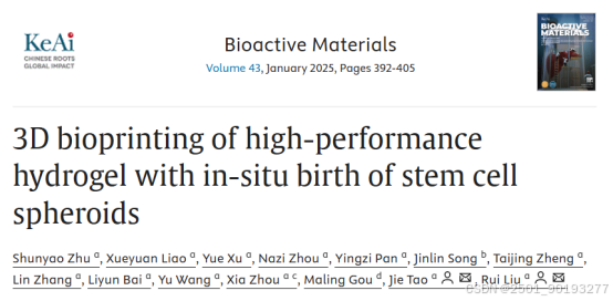
首先,研究者们观察到形成水凝胶构建体后,所有混合细胞都集中在葡聚糖溶液中,而在GelMA溶液中没有可检测到的信号。他们也发现CCB水凝胶固化后,超过80%的葡聚糖可以在24小时内,通过浸泡在PBS溶液中被有效消除。CCK-8测定结果表明,消除葡聚糖后的多孔结构促进了细胞的增殖。最终10%(w/v)葡聚糖溶液与15%(w/v)GelMA溶液按1:2体积比混合用于后续打印(图1)。
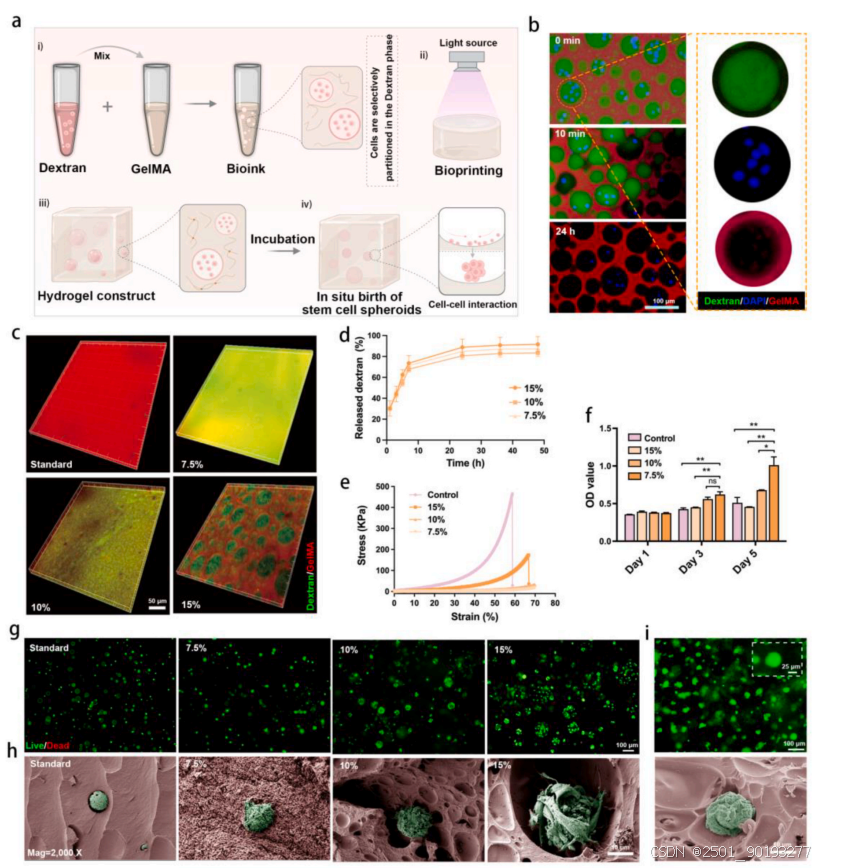
图1
Fig. 1. Characterizations of the bioink. (a) Schematic representation of the bioink used for DLP-based bioprinting and its application in facilitating in situ birth of stem cell spheroids. (b) Cell distribution and dextran removal process within hydrogels. (c) Visualization of different GelMA concentration with dextran at a volume ratio of 2:1 on the distribution of dextran. (d) The profile of released dextran. (e) The representative compressive stress-strain curve of the hydrogels. (f) Quantitative cell proliferation results of NIH/3T3 at day 1, 3 and 5 (mean ± SD, n = 3, two-way ANOVA). (g) Representative live/dead images of the NIH/3T3 cells at day 5. Live cells were stained in green, while dead cells were stained in red. (h) SEM micrographs of NIH/3T3 cells within hydrogel at day 5 cultured. (i) Representative live/ dead and SEM images of the BMSCs in CCB group of 15 %. Ns was determined as P > 0.05 with no statistical difference, *P < 0.05, **P < 0.01.
紧接着,研究者们进行了打印性能的表征,他们发现这一墨水配方可打印多种结构(从2D到复杂3D),负载细胞后的生物墨水也可实现精确沉积,表明这一生物墨水能实现高精度结构的制备(图2)。
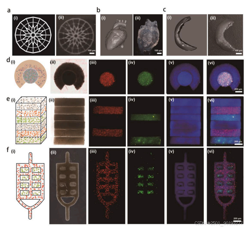
图2
Fig. 2. Printability of the bioink using DLP-based bioprinter. (a–c) 3D-printed wheel-shaped, heart-shaped and meniscus structure. (d–f) 3D bioprinting of hydrogels with different tissue models, including glioblastoma, layered skin and prevascularized hepatic hydrogel models, respectively
随后,研究者们利用大鼠牙髓干细胞(rDPSC)验证生物墨水是否可以促进干细胞球体的原位生成。结果显示,封装的rDPSC在CCB水凝胶中聚集形成球状体的能力显著上升,同时也表现出增殖能力的提高,干细胞标记物例如Nanog、Oct4和Sox2表达上调,预示其在维持细胞干性方面也具有一定作用。进一步,研究者通过转录组测序分析表明CCB水凝胶促进了多种生物过程和分子功能富集,例如细胞黏附、细胞间连接和细胞外基质形成等,有利于组织再生。同时,CCB水凝胶还能通过增加成骨、成软骨、成脂分化相关基因表达。促进rDPSC的分化能力(图3、4)。
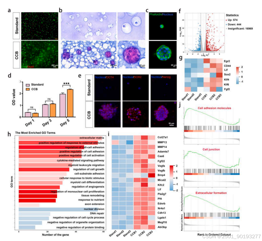
图3
Fig. 3. 3D bioprinting of hydrogel constructs modulated the activity of encapsulated stem cells and facilitated the formation of MSCs spheroids in-situ. (a) Fluo rescence micrographs showing the viability of encapsulated rDPSCs at day 5. Live cells were stained in green, while dead cells were stained in red. (b) Representative images of H&E staining of the rDPSC spheroids in hydrogels. (c) Cytoskeletal spreading in rDPSCs within hydrogel at day 5. (d) Quantitative cell proliferation results at day 1, 3, and 5 (mean ± SD, n = 3, two-way ANOVA). (e) Immunofluorescence staining of Nanog, Oct4 and Sox2 in rDPSC spheroids. Nuclei were stained with DAPI (blue). (f) Volcano plot of differentially expressed genes between these two hydrogel groups after 5 days of incubation. The red points represent the upregulated genes in CCB group. The blue points represent the down-regulated genes in CCB group. (g) Heatmaps of differentially expressed genes associated with stemness between two hydrogel groups. (h) Gene ontology (GO) enrichment analyses of upregulated and down regulated differentially expressed genes for total biological processes. (i) Heatmaps of differentially expressed genes associated with adhesion, extracellular matrix organization and angiogenesis. (j) Gene set enrichment analysis (GSEA) plot showing enrichment of the “Cell adhesion molecules”, “Cell junction”, and “Extracellular formation” gene set in these two hydrogel groups. The top portion of the plot shows the enrichment score (green line). The middle portion of the plot shows the appearances of the targeted genes in this gene set. The bottom portion of the plot shows the values of the ranking metric moving down the list of the ranked genes. Ns was determined as P > 0.05 with no statistical difference, ***P < 0.001.
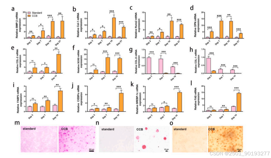
图4
Fig. 4. Multilineage differentiation of rDPSCs in hydrogel. (a–d) Quantification of gene expression of BMP2, Col-1, RUNX2, and OPN (mean ± SD, n = 3, two-way ANOVA). (e–h) Expression levels of cartilage-specific genes, including Col-2, SOX9, Col-x, and Col-1 (mean ± SD, n = 3, two-way ANOVA). (i–l) Expression levels of adipogenic-specific genes, including FABP3, c/EBPα, SREBP-1c and FAS (mean ± SD, n = 3, two-way ANOVA). (m) Optical microscope images of Alizarin Red S staining. (n) Safranin O-staining of the rDPSCs encapsulated in hydrogels. (o) Oil Red O staining of the rDPSCs encapsulated in hydrogels. Ns was determined as P > 0.05 with no statistical difference, *P < 0.05, **P < 0.01, ***P < 0.001.
另一方面,研究者们还利用人脐静脉内皮细胞(HUVEC)验证了CCB水凝胶构建体的血管生成潜力。结果显示,CCB水凝胶构建物促进了HUVEC的血管形成、节点数量、迁移速度。同时,将水凝胶构建体植入大鼠皮下,2周后的切片染色结果显示CCB水凝胶在血管密度、平均血管直径和管腔密度方面均优于对照组(图5)。
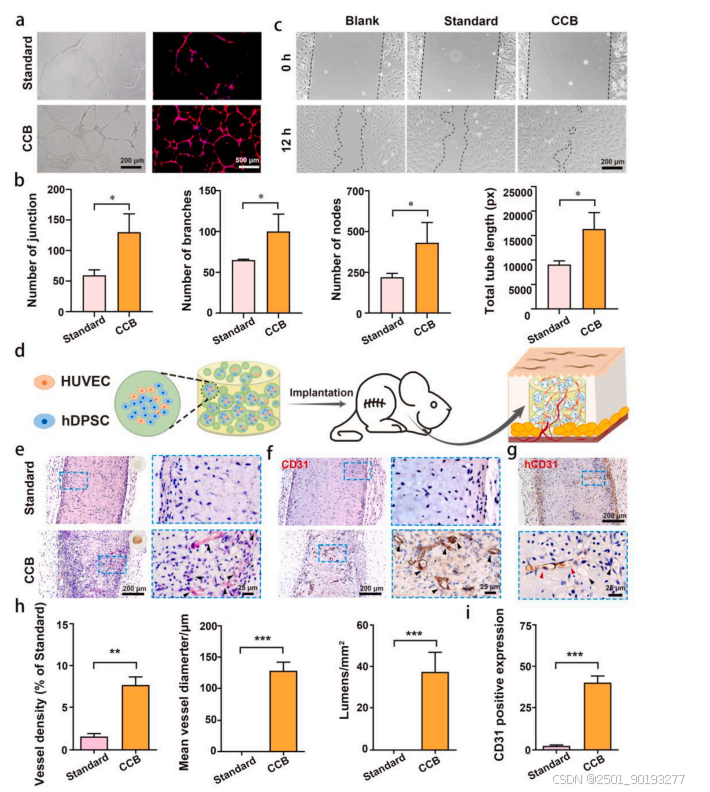
图5
Fig. 5. Application of bioprinted hydrogels in neovascularization. (a) Bright-field (left) and immunofluorescent (right) images of HUVECs in different conditioned mediums. (b) Corresponding qualitative analysis of vascular network organization indexes, including branches, junctions, nodes, and total length. (c) Wound-healing assays of the treated HUVECs with different conditioned mediums. (d) Schematic diagram of vascular regeneration in vivo. (e) Representative H&E images of angiogenesis in vivo. (f) Immunostaining images for CD31 at 14 days post-implantation. (g) Immunostaining images for hCD31 at 14 days post-implantation in CCB group. (h) Quantification of the vessel density, mean vessel diameter and lumens density from (e) (mean ± SD, n = 3, two-way ANOVA). (i) Quantification analysis of CD31 expression in (f) (mean ± SD, n = 3, two-way ANOVA). **P < 0.01, ***P < 0.001.
最后,研究者将3D打印的CCB水凝胶构建体填充于大鼠源性牙本质基质(TDM)根片并植入大鼠皮下用于牙髓感染治疗。体内标本组织学评估结果显示,8周时,CCB组中的TDM支架充满了新形成的组织,包括血管的出现和牙本质样结构的发育。此外,通过Masson三色染色观察到,在CCB组中有大量的胶原纤维。免疫组织化学结果显示,CCB组中DSPP和CD31的表达增加,证实了使用CCB水凝胶有效促进了牙髓-牙本质复合物样组织重建(图6)。
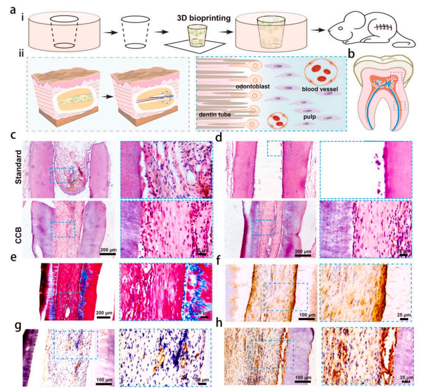
图6
Fig. 6. Reconstitution of dental pulpo-like tissue in a subcutaneous model. (a) Schematic diagram showing the i) fabrication of hydrogel constructs with TDM and ii) their application in regenerative endodontic treatment. (b) Schematic diagram of pulp regeneration sampling, with red boxes indicating border and green boxes indicating the center. (c and d) H&E staining of regenerated pulpo dentin tissue at different sites. (e) Masson’s Trichrome staining of regenerated pulpo dentin tissue in CCB group. Immunohistological staining of (f) dentin marker (DSPP), (g) blood vessel marker (CD31), and (h) nerve tissue marker (NF200) in CCB group.
综上所述:该研究中开发的CCB水凝胶生物墨水适用于基于DLP的组织构建生物打印,可促进干细胞球原位形成,制备的水凝胶构建物性能高、细胞活力好、结构保真度高,能增强干细胞特性维持和分化潜力,支持体内血管化和再生牙本质形成,为组织工程和再生医学提供新策略。
该研究由来自陆军军医大学口腔科的刘锐教授团队完成,并于2024年9月发表于Bioactive Materials期刊。
论文信息: Shunyao Zhu, Xueyuan Liao, Yue Xu, Nazi Zhou, Yingzi Pan, Jinlin Song, Taijing Zheng, Lin Zhang, Liyun Bai, Yu Wang, Xia Zhou, Maling Gou, Jie Tao**, Rui Liu*, 3D bioprinting of high-performance hydrogel with in-situ birth of stem cell spheroids, Bioact Mater 2024, 43: 392-405.
文章来源:https://www.sciencedirect.com/science/article/pii/S2452199X24004365?via%3Dihub
免责声明:本号对所有原创、转载文章陈述与观点均保持中立,内容仅供读者学习和交流。文章、图片等版权归原作者享有,如有侵权,请留言联系更正或删除。


 被折叠的 条评论
为什么被折叠?
被折叠的 条评论
为什么被折叠?
 到【灌水乐园】发言


