


近年来,得益于方法学的重大进步和从分子到整个大脑多层次的数字数据集成及建模,脑科学研究无疑已迈入一个新时代。在这一背景下,神经科学与技术、计算的交叉领域已取得重要进展。新兴的大脑科学整合了高质量的研究、多层次数据的集成、跨学科的大规模合作文化,同时促进了科研成果的应用转化。就如欧洲人脑计划(HBP)所提倡的那样,采取系统化的方法对于应对未来十年内的医学与技术挑战至关重要。
本文旨在为未来十年的数字大脑研究发展一套新概念,并与广泛的研究社区展开讨论,寻找共识点,以此确立科学的共同目标。同时,提供一个科学框架,支持当前及未来的EBRAINS研究基础设施发展(EBRAINS是HBP工作产生的研究基础设施)。此外,本文还旨在向利益相关者、资助组织和研究机构传达未来数字大脑研究的信息,吸引他们的参与;探讨综合性大脑模型在人工智能,包括机器学习和深度学习方面的变革潜力;并概述一个包含反思、对话及社会参与的协作研究方法,以应对伦理与社会的机会与挑战,作为未来神经科学研究的一部分。(本文为论文下篇,上篇参见:神经科学,跨越140年的创新与挑战 | 追问观察)
关键词:人类大脑,数字研究工具,研究路线图,大脑模型,数据共享,研究平台
▷Amunts, Katrin, et al. "The coming decade of digital brain research: A vision for neuroscience at the intersection of technology and computing." Imaging Neuroscience 2 (2024): 1-35.

脑科学的全球化
自21世纪初以来,脑科学研究领域数字技术的应用迅速扩展,现在我们可以分析来自数以千计大脑的多模态数据。这些数据通过公开的公共存储库(如英国生物银行)或全球网络(如 ENIGMA, HCP)提供。当然,如果不能将这些海量数据转化为知识,进而深入理解大脑的复杂机制及其在正常行为、成长、衰老及脑疾病中的作用,光有数据也是不够的。
因此,我们见证了复杂生成模型的兴起,这些模型结合了遗传信息和表型信息,跨越不同时间点来跟踪大脑状态的时空变化(Iturria-Medina et al., 2018; Vogel et al., 2021; Young et al., 2018)。人工智能策略在将庞大数据集分类为合理定义的子组中起着越来越重要的作用,这些子组可能适用于定制解读,例如行为倾向的多基因风险评分或药物临床试验的分层。这些方法最终为个性化管理或医疗干预提供了可能性。
然而,寻找更细微、更早期的大脑状态变化的生物标志物,常常需要汇聚大量数据来揭示那些与这些变化相关或可能导致这些变化的因素。这种搜索伴随着同质性与代表性之间的常见冲突。虽然毋庸置疑,大数据手段应用于广泛的公共数据存储库,如ADNI,PPMI,UK Biobank等,已经为我们提供了关于人类大脑机制和回路的通用性质的前所未有的洞察,但这些数据集大多源自西方国家,并不代表全球。
数据存储库的效用需要足够丰富和多元的数据,以确保研究成果及其推动的创新可以在全球范围内的多样化人群和环境中得到推广。性别差异、年龄、社会经济地位、种族等因素在神经结构、功能和认知表现上造成了个体差异(Dotson & Duarte, 2020),也影响了不同人群间疾病的发生率、康复和生存率的差异(Sterling et al.,2022; Zahodne et al.,2015)。此外,全球范围内关于研究中报告种族人口信息的做法存在差异(Goldfarb & Brown, 2022)。同时,低收入及中等收入国家(LMICs)在脑疾病和心理健康问题的诊断和发病率等方面的举措不断增加,如东南亚国家联盟(ASEAN)地区。
全球合作的需求包括收集、传播和分析来自LMICs的经过精心管理、详细表型和基因分析的数据集,以辨识全球不同亚人群间的相似性和差异性。在没有获取不同国家具有代表性的数据的情况下,无法对这些比较进行统计上的可靠推断,这超出了个别实验室的能力。由于对现有数据集的重复使用导致它们的不可避免的衰减(Thompson et al., 2020),代表性问题不能仅作为事后考虑,而需成为紧急的优先事项。
▷图源:Script & Seal
在接下来的十年里,随着开放数据共享倡议(如英国生物银行,OpenNeuro,CONP, EBRAINS等)在全球的扩展,科学家对数据管理和共享的观念将持续演变(Donaldson& Koepke, 2022),资助者和学术期刊的期望也将发生变化(可参见2023年Nature Neuroscience社论“我们如何促进数据共享”),这将极大地增加全球社区可用的多样化数据量。这将带来对相关和因果因素的新的认识,这些因素导致全球人群中大脑和行为差异的出现。这些数据共享平台,很多已经运行十多年,已经达到了技术上的成熟,能够支持多国之间的开放数据共享。
然而,在不同平台之间开发清晰而无缝的互操作性仍有待完成,以确保终端用户可以在不需要深入理解复杂技术细节的情况下进行操作。挑战不仅仅在于提供数据,更重要的是提供既有价值又易于解释的数据,这些数据的来源必须遵循FAIR数据共享原则(可查找的、可访问的、可互操作的、可重用的,Wilkinson et al., 2016)。从技术上实现数据互操作性、提供数据描述符和协议、遵守元数据标准,这些措施不仅提升了数据的价值和实用性,还有助于构建一个更强大、更协作、更高效的研究生态系统。
然而,获取有意义和可操作数据的必要性,也带来了一系列与数据治理和伦理相关的挑战。这些实践在不同群体间仍在演变,拥有多样且有时不兼容的全球框架(Eke et al., 2022)。关于研究中种族人口信息的报告也存在差异(Goldfarb & Brown, 2022),以及生成和处理数据的技术能力、数据收集的资金和其他社会文化因素也是考量因素。到目前为止,来自非洲和拉丁美洲地区的数据集通常不被包括在全球脑科学研究和创新的讨论中。
下一个十年将见证在欧洲(如GDPR)、北美、亚洲、澳大利亚和非洲等地区不同的数据治理和伦理框架的协调,以促进大脑数据在开放神经科学全球社区内的更广泛传播。我们应更加关注能力建设、增加人口信息的报告、资助计划,并最终提高低收入和中等收入国家对数据生成、处理和分享的意识。
毫无疑问,脑科学全球化的最重要的内容将是其“民主化”。不再是仅仅由高收入国家的科学家分析和发布的数据来源,我们预计LMIC的科学家将在脑科学事业中扮演越来越重要的角色。此种民主化自然演化自当前数据分析门户(如CBRAIN、EBRAINS、BrainLife*)所提供的高级分析工作流程的普及。这些门户允许来自世界各地的研究者在其他地方进行复杂的数据分析,消除了后勤、行政和技术障碍,这些障碍曾经阻碍LMIC的科学家充分参与到脑科学社区。此外,通过结合数据共享和分析平台,还可以实现派生数据的重新分配。共享结果至关重要,能够最大限度地减少科学冗余、增强可重复性,并促进LMIC场景中科学分析的可访问性。
CBRAIN:https://cbrain.ca/;EBRAINS:https://ebrains.eu/;BrainLife:https://brainlife.io/
随着人们对分析决策在学习大脑模型中的作用认识的增强(Botvinik-Nezer et al., 2020),派生数据的传播将使科学探索的迭代和协作方法成为可能,并消除了进入的主要障碍。这种愿景也带来了需要解决的一系列行政问题,例如学术认可、晋升、指导等,但这些问题已经是当前开放神经科学辩论的主题。全球化的拓展带来了规模与后勤的挑战,例如语言和地方治理法规的问题,但这并不改变数据隐私与开放科学之间基本的矛盾。我们预期随着技术挑战的解决,全球神经科学整合的愿景将在未来十年成为现实。

大脑模型作为未来脑研究的推动力
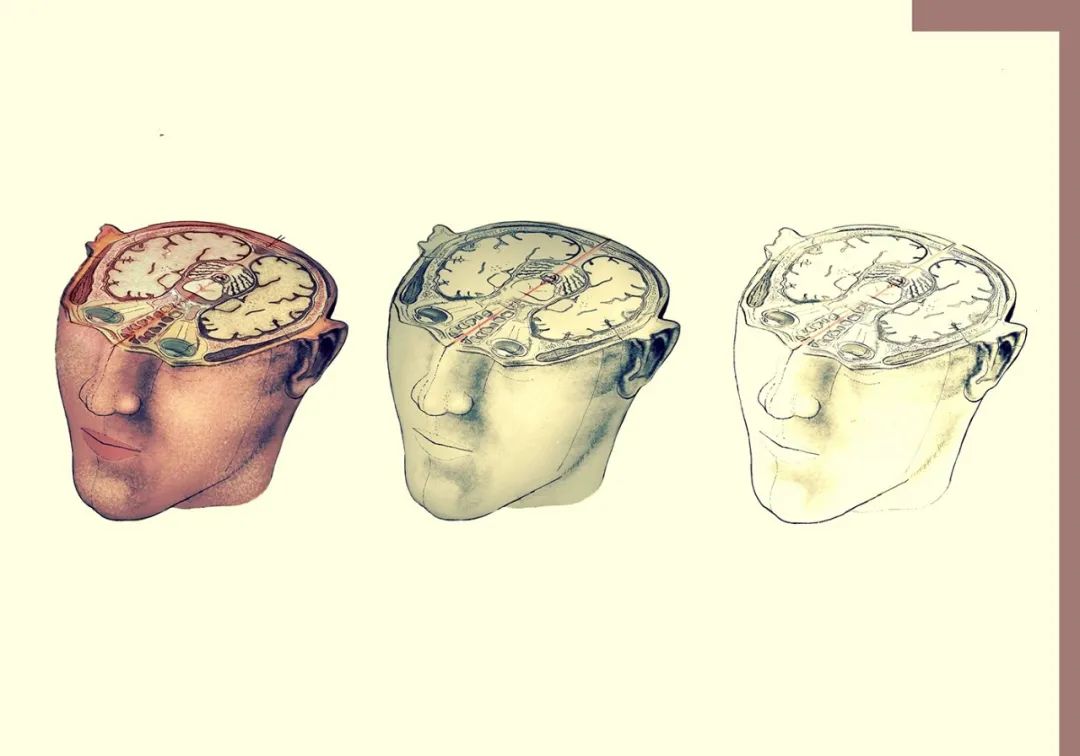
在过去二十年里,信息和通信技术的迅猛发展不仅推动了模拟和机器学习技术的进步,也使得数据与模型在同一生态系统中实现互联互通,从而推动了新型脑模型的发展。大脑模拟直接利用了大脑基础研究的成果,预计将在阐释脑过程的基本方面(通过展示其在体外模拟的能力)如决策制定、感觉运动整合、记忆形成等方面发挥关键作用。尽管我们需警惕这些研究所带来的伦理与哲学问题,但也可以设想利用这些模型和模拟来探索脑研究中的具体问题。由此,我们不难设想如何定制通用脑模型,以捕获某一特定患者大脑的独特特征。例如,个体的结构和功能性脑成像数据可以约束一个通用的数字脑模型,使其针对特定个体,从而用作个性化分析模板或体外模拟平台。
这种方法的一个具体例子是虚拟癫痫患者,在此方法中,神经影像数据指导对癫痫患者大脑的体外模拟,支持诊断和治疗干预、临床决策和后果预测(El Houssaini et al., 2020; Jirsa et al., 2017; Wendling, 2008)。在计算神经科学的总体趋势下,基于相关神经回路知识,各种癫痫活动模型被构建。这些模型通常将神经元或神经群体网络的癫痫发作解释为一种高同步性/高振幅节律状态。在无法直接从受试者获取数据的情况下,多级图谱数据成为另一种可以进一步丰富个性化脑模型的数据来源(Amunts et al., 2022)。
这些个性化的“虚拟大脑”可以被看作是向理论和技术上更具挑战性的新阶段迈进的一种跳板,这些挑战在伦理方面可能更为复杂,同时也更适应于大脑活动在所有时间尺度上的不断变化。个性化脑模拟的终极目标可以体现在一个连续通过真实世界数据得到信息和更新的模型,这种模型被称为“数字孪生”。在这一背景下,“数字孪生”的概念需要被仔细界定,以避免掩盖这种方法的局限性,并避免制造对精确度的不切实际期待或产生适得其反的过度宣传(Evers & Salles, 2021)。
历史上,“数字孪生”的概念起源于工业和制造领域(Grieves & Vickers, 2017; Grieves, 2019),包括三个组成部分:物理对象、其虚拟对应物和两者之间的数据流动。物理对象的实测数据传递给模型,而模型的信息和过程反馈给物理对象。今天,“数字孪生”一词已经广泛应用于其起源之外的多个研究领域,包括生物医学领域,尽管该术语背后的概念可能存在差异。
在制造业中,数字孪生不仅仅是一个普通的模拟模型。它是为特定对象制定的通用模型的具体实例,由该对象的实际数据支持,例如在工业领域中的飞机引擎(Tao et al., 2019)。最近,在相同的背景下,研究者还提出了“数字影子”这一概念作为一种改进方法。这种方法提供任务和情境依赖的、目标导向的、聚合的、持久的数据集,能以更灵活的方式涵盖多个视角下的复杂现实,并且性能超过完全集成的数字孪生(Becker et al., 2021; Brauner et al., 2022)。
▷图源:Matt Chinworth
数字孪生的一种解读涉及到机器学习和人工智能中生成模型的辩证关系。生成模型保证了模型的可解释性。此外,它们促使我们从“大数据”向“智能数据”的转变(更确切地说是选择和整合数据特征,以最大化预期的信息增益)。生成模型是从潜在原因到可测量结果的映射的概率描述。在这个意义上,数字孪生可以看作是一个适合生成某个特定细胞、个体或群体反应的模型的正式定义。正确构建生成模型关键在于,它能够提供对实验数据的可解释的机械性解释。此外,它分别在模型拟合(即反演)和模型选择(即假设)方面区分了自下而上与自上而下的建模方法。
在构建一个活体器官的“数字孪生”时,面临的挑战超出了构建一个无生命对象的数字孪生时的挑战。大脑无疑是目前已知的最复杂和多面的器官。那么,在神经科学和大脑研究中,数字孪生的概念能够被多大程度地应用呢?如果简单地将数字孪生概念1:1地应用于大脑,可能会引起严重的误解。在这里,我们希望通过在脑科学的特定背景下明确定义这一术语,为相关讨论做出贡献。我们区分了目标驱动的数字孪生和大脑的完全数字复制品(或副本/复制),后者代表了大脑所有层面所有方面的完整呈现(参见Box 3)。
大脑的完全复制既不可实现,也似乎没有明确的实用价值。我们讨论中的数字孪生应被理解为一个虚拟模型,旨在充分代表一个对象或过程,受其物理对应物的数据约束,并提供模拟数据以指导选择并预见其后果。数字孪生因此是实用意义上的复制,通常与一个功能或过程的模型相关,其力量在于它在处理其物理对应物所面临的相关问题时的有效性,保持适当的抽象水平。因此,其目标不是尽可能地详细和多层次地模拟生物大脑,而是选择性地减少那些对特定研究问题具有预测价值的数据信息量,保持模型尽可能简单,同时确保其复杂度足以应对需要。
即便是专门用于理解特定大脑结构和动力学,或是预测特定患者的病情进展的模型,也需要依赖于全面而复杂的数据源,以构建信息丰富的虚拟大脑模型。例如,人类大脑计划已在EBRAINS上建立了一个高分辨率的多层次人类大脑图谱,作为结构与功能数据的集成平台。对于每个模型,我们都需要证实增加的数据是否真的增强了模型的强度,即这些数据是否使预测更准确、可验证?我们需要持续监控在更好的预测与收集数据的可行性及相关成本之间的权衡,并评估这些数据选择是否适合当前的问题,即是否反映了关键的决定因素(Box 3)。
Box 3:数字大脑模型分类
大脑模型:大脑模型是大脑的数字表示,这一术语在不同的情境中有不同的用途;常见的包括数字图谱、人工神经网络、解剖模型、生物物理模型、网络模型、认知和行为模型,以及数学和数据驱动的模型。
个性化大脑模型:个性化大脑模型是一种特殊类型的模型,通过将一个个体的特定数据整合到更广泛的模型中来进行个性化(例如,通过虚拟癫痫患者实现)。
数字孪生:下一代个性化大脑模型,它们通过不断地融入实时数据而不断发展。这些模型是为了解决特定研究问题而有目的地设计的,整合了相关的数据。
完全复制:这是一个假设的概念,指的是在所有层面上完整地数字化表示一个大脑的想法,最终包括对数字孪生体的解释。
数字孪生与其他个性化虚拟大脑模型的一个显著区别在于,数字孪生能持续接收来自现实世界的新信息,以实时适应其环境。在神经科学领域,大脑的“数字孪生”极具前景,可用于持续调整功能性神经康复的干预措施或定制神经技术干预方案。应用高保真的准实时更新的人脑数字孪生模型,需要在技术上进行开发,如将孪生大脑生态地沉浸于模拟环境、高带宽稳定的脑机接口和极高的计算能力等,这些领域的突破仍是遥远的长期目标。尽管如此,数字孪生已在神经科学和医学领域找到应用,前提是充分考虑到当前大脑模型的局限、个性化过程及技术更新频率的挑战。数字孪生定义了当前数字神经科学发展路径的视野,并应被视为未来发展的驱动力。
尽管大脑的数字孪生在具体应用上还有一段距离,但数字大脑研究的时代已经无疑开始了,无论是在现实世界还是在研究领域都是如此。数字大脑研究是一个综合概念,涵盖了数据、模型、理论、方法和计算技术,集成于 HBP 框架下的所有研究和开发工作。它的价值体现在成功演示内部和外部有效性、生态和构建有效性等方面。这使研究人员能够应对神经科学数十年来面临的主要挑战,如个体内外变异性、机制不明确性和多尺度复杂性等问题。EBRAINS 提供了一个平台和用户界面,支持数据、模型和方法组件的互操作性,为数字大脑概念在神经科学研究中占据中心舞台提供了操作基础。
我们认为,在短至中期内,数字大脑模型可以在以下三个领域发挥重要作用:(1)基础大脑研究,(2)医学应用,(3)基于大脑的技术开发。
基础大脑研究
数字大脑模型及其模拟并不会替代基础研究和知识积累,而应视为一种有益的“工程”工具。它目前充当一个在进展中的预测模型,旨在(1)检验现有知识,(2)预测干预效果。后者尤为引人关注,因为干预手段正不断增多,诸如深部脑刺激(DBS)、经颅磁刺激(TMS)、经颅直流电刺激(tDCS)、经颅聚焦超声刺激(tFUS)、药物、光遗传学和光药理学等。虽然已有多项研究利用计算大脑模型来进行预测、指导干预研究的设计并解释观测到的效果(Frank et al.,2004,2007),但这些方法目前往往是基于“半经验”的应用,涉及电极位置、电路连接、功能及电气模型、神经元类型的遗传启动子、神经受体的表达模式及其信号通路模型等信息。数字孪生技术可能促进这些参数的合理决策,测试结果,并随后对模型进行评估和修正等。
为了取得成功,底层模型必须具有生物现实性,即在解剖上精确且在功能上全面。它们最终应能关联大脑结构与功能和行为,并可能用于研究认知、语言、意识或情感。这需要整合不同层次的高度异质数据,包括体内和离体数据,并将它们置于相同的空间参考框架中。在一种替代而互补的方法中,细胞图谱网络(BICAN)将采用美国细胞普查网络(BICCN)的方法,扩展至整个人脑,对哺乳动物大脑的组成部分进行深入的特征描述,例如,对初级运动皮层的最详尽、最全面的多模态模型进行研究,这包括单细胞转录组和蛋白质组、染色质可及性、DNA甲基化组、空间分辨单细胞转录组、形态和电生理特性及细胞分辨率输入输出映射(Callaway et al.,2021)。
基于这一概念,大脑模拟在阐释大脑的复杂性中扮演了关键角色,它通过允许测试关于大脑多级组织及其控制周围身体功能的假设来实现(参见下文)。显然,沿此研究方向,不同空间层面上执行的模拟的相互连接将变得日益重要。例如,分子层面的 EBRAINS 模拟引擎 Gromacs、细胞层面的 Arbor 和 NEURON、系统层面的 NEST、全脑层面的 Virtual Brain 以及体现生物体及其环境的神经机器人平台(见 Brain-derived technologies);概述见 Einevoll et al., 2019。
与真实活体大脑不同,嵌入式模拟大脑可以在任何空间和时间点进行抽样。因此,我们能够监测到模拟大脑中所有基于现实世界数据或物理化学模拟的过程,并使用模拟测量设备如多阵列电极、fMRI扫描仪来观察。理论上,它可以在全身闭环环境中测试各种功能假设;此外,还可能构建动力学解剖图谱,例如在特定刺激下观察大脑区域的变化和过程的图谱,所有这些都能在真实模拟的实时中实现。
活体大脑的复杂多尺度结构、有限的测量可接近性和对大脑过程理解的不完整,使得数字孪生技术的实施极具挑战。BigBrain 作为一个解剖模型可能成为严格意义上整合孪生数据的支架(Amunts et al., 2013),这些数据包括其他来源的动力学细胞数据、实验人群研究的数据以及由模型和不同大脑模拟的合成数据。这种方法也定义了数字孪生策略的限制和有效范围,对于负责任地使用此技术及其后续的信任至关重要。然而,这些数据驱动的模型可能代表了在任何特定时间点可实现的活人大脑的最接近的数字表示。未来,数学的新见解将必要地加速模拟和模型分析(Lehtimäki et al.,2017,2019,2020)。
据此,我们可以设定以下目标:(1)发展多层大脑图谱和高分辨率的大脑模型。(2)启用多层大脑模型和模拟。(3)揭示认知和行为的机制。
▷图源:Art and Science
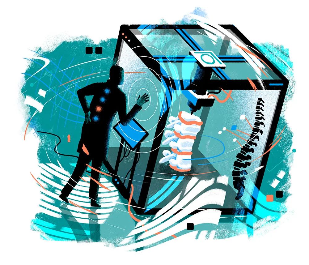
大脑医学
从这些数字孪生技术中,我们可以衍生出个性化孪生技术,目的是以全新且高效的方式改善患者的诊断和治疗,支持大脑健康的战略,正如欧洲神经学院最近发布的相关策略所示 (Bassetti, 2022)。与心脏数字孪生相似 (Gillette et al., 2021),即基于临床数据生成的与所有可用临床观察数据相匹配的患者心脏数字副本,人类的电生理副本在指导临床决策方面显示出巨大潜力,并且有助于以成本效益高、安全且符合伦理的方式测试新的设备治疗方案。医学中的数字孪生专注于特定的空间规模,具有明确的粒度,涵盖特定的时间间隔,服务于特定的目的。近期提出了针对阿尔茨海默病的数字孪生方法 (Stefanovski et al., 2021),尽管需要谨慎考虑数据隐私、安全性和安全方面的问题,但个性化孪生也可能成为治疗此类疾病的一个非常有力的策略。
虚拟大脑(Virtual BigBrain,TVB)允许根据受试者的神经影像和 EEG 数据以及 BigBrain 模型的解剖数据构建个体化的连接组 (Jirsa et al., 2017)。正在进行的EPINOV临床试验采用了 TVB,这在该领域是一大进步;科学家们开发了患者脑部的个体模型,以指导和预测癫痫手术的最佳治疗效果 (Jirsa et al., 2023; Proix et al., 2017; Wang et al., 2023)。他们所用的策略是将群体数据与个别脑部数据结合,开发出足够真实的虚拟脑模型,也就是孪生体,使得可以在手术前进行干预模拟。对于那些在麻醉期间仍持续发作的难治性癫痫患者,通常需要长期的重症监护,并面临极高的永久神经损伤和死亡风险。对这些患者而言,数字孪生可以用来审查大量模型,持续获得来自 EEG 的反馈、药物反应以及血液中离子和气体的浓度等信息,这些都是重症监护环境中容易获取的数据。
数字大脑建模的实用性由DBS证实,DBS是几种难治性神经疾病的成熟外科治疗方法。目前,临床上的 DBS 通常采用“开环”系统,即按照固定参数持续施加刺激。这些参数在植入后可调整,但调整是手动进行的,且操作不频繁,主要基于观察患者的明显症状。相对而言,“闭环”、自适应的DBS被开发出来以克服传统DBS的限制,它根据实时的临床相关生物反馈信号调节神经回路 (Marceglia et al., 2021)。然而,成功应用这些技术,需要深入理解神经可塑性和学习机制。
面对局部大脑损伤如中风或创伤性脑损伤的应用也需类似的技术。除了侵入性治疗干预,数字孪生也是一个预测大脑损伤后果、病理生理和可塑性的强大工具,有时这些可通过计算神经心理学来描述,即使用合成损伤在计算模型中模拟损伤与缺陷之间的关系 (Parr et al., 2018)。这可以显著提升我们个性化神经康复的能力,同时整合由虚拟现实和机器人治疗产生的复杂信息,以及精确测量患者的反应和进步。
其他应用可以利用模拟测试一个规模远大于真实人群的“临床”模拟人群,从而通过创建“数字患者”群体来放大数据。这种方法对于评估罕见病、研究性别影响或预测疾病进程尤其有吸引力 (Maestú et al., 2021)。此外,使用的数据源越多样和异质,模型在其他数据集上的表现就越好,这也提高了模型的普适性。这是联合系统提供的一大特色,它有助于增加数据来源的多样性(例如,Dayan et al., 2021)。
最近,DeepMind 开发的 AlphaFold 系统 (Jumper et al., 2021),该系统通过应用深度学习方法,已能够预测蛋白质的 3D 结构。这种技术可推广至系统级,用于测试药物与蛋白或药物-蛋白系统的相互作用。此外,从在虚拟环境中测试药物的效果到揭示药物在分子及系统级别的作用机制,这些都是此技术的进一步发展方向。考虑到量子力学/分子力学在计算上的高要求,这种系统级的方法需要在最强大的超级计算机上运行的高度可扩展工具。可以使用NEURON和Arbor构建和模拟的精细的局部微电路模型,直接用于映射某些分子(如离子通道、受体)的局部分布,然后用来模拟药物对这一系统的影响。这些小规模模型可以根据特定病理条件进行调整,然后转化为针对患者的平均场模型,提高数字孪生的精度。
更广泛地说,人类大脑研究领域与非人类大脑研究领域的增强交流,可能会协同解决生物医学科学中长期存在的问题 (Devinsky et al., 2018)。人类和伴侣动物患有一些相同的疾病(例如癫痫、癌症、肥胖)。像人类一样,患有癫痫的狗在生病时也需接受脑部扫描。这些疾病和治疗的重叠表明,人类医学和兽医学之间存在未被充分利用的机会,这些机会可以用于在伴侣动物中测试个性化医学和数字孪生的有效性,进而在人类中实施。
最后,大脑孪生技术预计将有助于发展“人体孪生”技术。这一视角超越了单纯增加一个器官的层面,因为它将允许在系统级别模拟神经系统活动与其他器官的相互作用,例如心脑耦合,以及大脑与胃肠道的连接。这些相互作用广泛且双向。例如,最近的研究发现,人类大脑中有一个固有的调节内环境和内感觉系统,包括控制身体内环境的皮层控制区域,支持身体的恒常性调节 (Kleckner et al., 2017)。此外,如呼吸等身体过程是节律性神经活动的重要推动力 (Tort et al., 2018)。捕捉这些双向互动将有助于我们理解大脑如何支持重要的身体功能——可能还包括在功能受损时如何恢复它们。
欧洲委员会目前正在制定的数字人孪生路线图中,多器官或多尺度数字孪生的双向和系统性链接是一个关键要素 (https://www.edith-csa.eu/)。
因此,研究者可以确定以下目标:(1)在生命周期中获得关于大脑可塑性、学习和适应的详细见解。(2)加速数字大脑医学的发展。(3)探索并模拟大脑作为身体一部分的模型。
▷图源:Mandara Nagaraj
大脑衍生技术
一项基本挑战在于确定大脑建模所需的精细度级别、过渡性计算以及模拟开发的类型,以便支持各种认知和感觉运动功能的涌现。模拟人类大脑的模型被设置在具体环境中,即这些模型能控制虚拟或实体的身体与现实的虚拟或实际的物理环境互动,并接收依时间变化的输入流来产生行为输出,这为研究大脑结构、大脑活动与认知及功能表现之间的联系提供了一个极具吸引力的平台。
如何评价这种自下而上的组合及数字孪生系统的涌现行为与生物数据的一致性,仍是一个持续的挑战,因为典型的合成发展环境与自然环境不一致。Yong (2019) 在《大西洋》[12]杂志的特稿《人类大脑项目未能兑现其承诺》(The Human Brain Project Hasn’t Lived Up to Its Promise)中指出,“大规模模拟有助于理解气象和星系,但行星系统只关注它们自身。而大脑则是为了处理其他事务而构建的……模拟组织是可行的,但没有意义。”
前文段落列举了几例模拟在基础神经科学和大脑医学中取得进展的例子,针对的是明确的研究问题。此外,从一开始,HBP便旨在发展技术,以便研究大脑与环境的互动(Booklet,2016)。换言之,某些大脑过程的模拟被嵌入到一个真实或模拟的身体中,其所有传感器和执行器都与模拟相连。原则上,这些传感器和执行器可以是真实的、模拟的,或两者的结合。同样,这个身体被置于一个真实或虚拟的世界中。一旦拥有了这些元素,无论是模拟的还是真实的,我们就能以任何合理的方式组合它们。
显然,这种方法高度依赖于模拟真实世界物理现象的模型,并且还需要复杂的软件来高保真地模拟空间环境,并提供足够的环境、传感器和执行器物理模拟,连接大脑模拟器,提供存储模拟结果的设施、图形渲染和这些复杂软件模块的协调。所有这些(共同)模拟可以在不同的时间尺度上运行(理想情况下当然是实时的),在闭环或开环的情景中,并且以不同的粒度对实体进行建模。
HBP 的神经机器人平台[13]是一个专为执行所有这些步骤而设计的软件环境,它基于来自生物实验的多样化数据集和真实世界机器人的输入运行模拟,并在这些模拟的基础上整合了机器学习。虽然这个平台最初是为设计那些由生物学启发的大脑模型控制的神经机器人而构思的,但它随着时间的推移已演变成一个能够连接和整合从模拟小鼠身体到复杂传感器模型,以及各种神经元和大脑模拟器的各种实体的软件环境。如今,神经机器人平台不仅是一个机器人设计的环境,同时也是执行神经科学实验的平台。因此,它是一个强大的虚拟神经科学工具,甚至可以用完全在该平台内运行的计算机实验取代系统级体内实验。
此外,神经机器人平台还允许在机器人建造之前,用真实的神经科学数据来训练具体化机器人的“大脑”(基于 AI 的控制器)。可以想象,一个模拟的真实环境副本可作为训练的基础,从而让机器人在被交付给终端用户之前进行预训练,用户只需对(涌现的)行为做出小的调整,以确保机器人能够完美执行其任务。我们将这种模式下的方法称为大脑衍生技术,因为它们直接基于并建立在大脑研究的发现之上。重要的是,这些发现可以在不同的组织层面得以实施。
在神经形态工程中,主要组件即生物神经元,通过功能等效的电路被模拟,构建高能效的模拟处理器和传感器。运行在这些系统上的神经模型可以源于已在生物大脑中识别的特定类型的神经元、微电路或大脑区域。当这些系统与机器人实体(无论是模拟的还是物理的)或生物体相连接时,它们可以复制感知、认知和行动的完整闭环的某些方面。因此,建模可以扩展到整个有机体,并覆盖复杂认知过程在行为层面的所有方面。大脑衍生技术因此不仅限于模仿大脑的结构特征,还可以包括认知模型和架构以及其基础的神经动力学。这些技术将成为大脑研究的新工具,并推动计算、机器人学和人工智能领域的创新。
神经康复领域预计将极大地受益于这种方法,其中现实的大脑-身体互动模型将有助于揭示发挥作用的神经机制(Rowald & Amft, 2022)。通过将详尽的大脑模型与脊髓和肌肉骨骼系统的模型结合,为我们提供了独特的机会,来详尽地研究神经活动与运动行为之间的关系。因此,个性化模型因此可以整合到决策支持系统中,帮助医生或治疗师选择和组合康复策略。它们还可能支持中央神经系统(包括脊髓)刺激技术和功能性电刺激的突破性发展,提高这些技术的效果并扩大它们的适用范围。最近一项成功的硬脊膜外电刺激治疗脊髓损伤的应用报道显示了这种方法的潜力(Rowald et al., 2022)。
同样,高保真的人体肌肉骨骼系统和中央神经系统模型的结合,有望支持所谓的电子药物(electroceuticals)的计算机技术的出现,这些设备用于治疗目的的医疗设备(例如,在帕金森病、癫痫等疾病中提供神经刺激)。医疗设备行业无疑对指导其产品设计、生成疗效预测以及整体降低产品开发过程的风险具有根本性的兴趣。因此,利用 HBP 创建的大脑图谱和多尺度大脑模拟器,似乎应该及时考虑收集和整合新数据(例如介电特性),作为开发用于评估电子药物的模拟工具和服务的前奏。考虑到DBS已被广泛使用,模拟这些电子药物的效果显然迫在眉睫。
▷图源:Valentin Tkach
HBP已支持 SpiNNaker 多核和 BrainScaleS 物理模拟神经形态计算平台建立首个开放的神经形态计算服务,并为这些技术的进一步发展做出了贡献(Furber & Bogdan, 2020)。神经形态技术,其中数据传输和处理都是基于事件的,即基于脉冲的,为边缘计算、移动机器人和神经义肢技术提供了多种机会。
考虑到移动系统自动化和“始终在线”传感器阵列的当前趋势,特别是神经形态设备有望提供增强的低延迟容量,用于感知、认知和行动,同时减少系统上操作对系统能源消耗的影响(Cramer et al., 2022; Göltz et al., 2021)。例如,将产生脉冲的处理单元与产生脉冲的传感器(例如,动态视觉传感器、动态音频传感器)结合成完整的神经形态系统,将使数据融合更加容易,并克服与数据来源异质性相关的限制。通过突触可塑性,尤其是神经回路学习的神经计算理解的进展,也将为赋予神经形态电路更复杂功能提供新的方法,并降低训练成本(例如,一次性和连续在线学习)。特别是,对局部可塑性的限制构成了相对于传统冯诺依曼架构的明显优势。
如 BrainScaleS 所示,模拟生物神经元的离子流动的模拟神经形态处理系统的电路是通过电流实现的。与基于经典冯·诺依曼架构的传统微处理器不同,每个硅神经元都被物理地嵌入到芯片中,配备专用组件。就像大脑中的神经元一样,这些硅神经元通过脉冲交换信息,这种方式极为高效,也是神经形态系统成为新一代实时且节能计算机的前景光明的原因之一。他们直接从大脑的结构派生的重要后果是,神经形态处理器通常不适合加载外部数据,而是支持实时在线学习。这种独特的功能使新类型的学习规则成为可能,这些规则不需要庞大的数据集,而是可以根据需要动态适应。
基于脉冲时序依赖性可塑性的学习规则是大脑衍生系统的一个显著成功案例(Diamond et al., 2019; Kreutzer et al., 2022)。它们直接植根于实验结果,并已成为理论神经科学和神经形态工程研究学习算法的基石。值得注意的是,传统机器学习也极大地受益于大脑研究。其中最著名的例子可能是卷积神经网络,其理念最初就是从视觉皮层的结构中提取而来的。
神经形态传感器是基础大脑研究促进新技术出现的另一个重要领域,尤其是动态视觉传感器和动态音频传感器。前者模仿视网膜的功能,并且像神经形态处理器一样,用尖峰编码信息。它们的特点与传统的同类产品完全不同。由于它们只发出变化信号而不是捕获完整图像帧,因此它们能以极高的效率运行,催生了新型图像处理算法,并理想地与神经形态处理器相结合。
从技术角度来看,人类大脑也被视为在人工系统中实现高级认知能力的最有前景的“罗塞塔石碑”。现代人工智能体的特点是智力水平有限,难以在提供的训练集之外进行泛化,其对环境的理解通常也较为肤浅。缺乏泛化能力意味着需要大数据集(资源密集型的大数据范式)、持续的人工监督(远程控制系统)或广泛且严格的任务规划以应对各种情况(如用于行星或海洋探索)。感知的肤浅和缺乏可解释性导致人工感知系统的鲁棒性和可靠性不足,这是实现有效的自动驾驶等技术的已知障碍之一。为了克服这些限制,必须开发与新的具身和增量学习算法相结合的大脑启发的多区域模型架构,以寻找最能模拟人类感知认知功能机制的那些算法。利用这些机制并理解认知功能的涌现将是创建可解释、可靠并最终更通用的人工智能的关键。
大脑的功能架构及其不同区域是为技术系统定义许多类型认知架构的基础。这对于机器人学尤其如此,其中大脑衍生方法被广泛研究。包括研究与具身相关的现象或开发新型感知和传感系统的例子,如受实际啮齿动物的体感系统启发的人造触须。
人工智能应用的神经网络未来的发展将看到主流人工智能与神经形态技术之间的融合。多尺度大脑模型可以为构建高级机器人控制器做出关键贡献。这些控制器可以嵌入塑性规则并通过与环境的互动自主适应。因此,基础大脑科学将是这些技术发展的关键信息来源。此外,神经形态计算可能有助于减少大型深度学习模型的大量碳足迹(Strubell et al., 2019)。
由此,可以推导出以下目标:(1)桥接人类与机器智能之间的差距。(2)构建神经形态大脑模型和仿生人工智能。

结论
要深入理解大脑功能,必须更加深入地了解大脑的组织结构以及基本的生物过程、它们之间的相互关系及其规则。这是提高预防、治疗及基于机制的诊断效率的基础。在未来十年的数字大脑研究中,一个有希望的方向是开发能够进行个性化模拟的大脑数字孪生体。虽然目前已可行,但大脑的数字孪生体仍处于初期阶段,开发完成后必须经过严格的测试和验证,才能有效应对大脑疾病,并成为颠覆性新型健康技术的基础。因此,我们需要理解系统及其子系统的计算目标和算法,以明确在个案实施中的限制和可能性。此外,大脑孪生体所引发的伦理问题需要我们与社会公开对话并加以解决。孪生体可视为大脑模型和分析持续发展的一个终点。
为实现这一目标,构建一个能够承载大脑数字孪生体的数字基础设施,有助于我们理解规则并改进数字大脑孪生体,直至通过验证测试,并可用于临床应用。此外,这种基础设施理想情况下应当提供互操作性、信息安全、多层次数据以及访问基于知识的计算资源,包括高性能计算和其他相关技术。EBRAINS 就是一个能承载这些发展的基础设施。要成功实现这一目标,对年轻一代进行培训,使其能够熟练利用这些基础设施和新的数字工具,显得尤为关键。
构建结构化数据和知识,以便研究社区能够轻松重新组合并集成,从而构建出众多的数字大脑孪生体,并提供执行这些孪生体复杂模拟的强大技术,这本身就可能成为一种颠覆性技术,帮助我们获得科学上的新洞见。
▷图源:Mark Conlan

科学目标:一份路线图
以下的“路线图”概述了未来十年内八个相交叉的研究领域的目标,涵盖了从近期或当前工作,中期,到长期的不同阶段。这是基于之前提供的输入得出的结论。
开发多层次大脑图谱和高分辨率大脑模型
近期:将从整个大脑到细胞的数据整合成一个全面、高分辨率的大脑图谱,作为深入理解大脑组织基本原则的基础,以预测图谱不完整部分的特征,并指导关于物种间相似性和差异的比较研究。
中期:制作详尽的、数据驱动的、多尺度模型,以研究人类大脑组织在不同生命阶段及不同条件下的变异性。
长期:阐释大脑组织和结构中负责复杂行为、智力和意识相关方面。
启用多层次大脑模型和模拟
近期:实现模型的多尺度整合,从局部生物物理属性到整个大脑模型,包括详尽的自下而上和自上而下的模型。这些模型将由数据及其预测测试驱动和调整。
中期:利用多尺度、全脑模型模拟生物学真实的复杂大脑功能,逐渐实现具体应用场景的数字大脑孪生。
长期:将模型预测应用于基础科学、医学和人工智能的大规模应用案例中,从而推动模型的测试和进一步完善,形成一个“生产性循环”。
阐明认知和行为的机制
近期:从多尺度角度出发(从感觉和视觉运动功能到更复杂的认知功能),建立描述认知功能机制的连贯框架。
中期:构建一个关于语言的连贯框架,作为人类独有的复杂认知功能,融合语言学和神经科学的研究洞见,通过研究发展过程窥探大脑专业化,并为语言模型和人工智能的发展提供基础。
长期:将各种假设下的概念和自我意识相互联系,并与细胞、分子及遗传层面的机制相结合。
在生命周期中获得
大脑可塑性、学习和适应的深入洞见
近期:识别可塑性、学习和适应的规则并将其整合到现有的多层次大脑模型中。
中期:确定大脑可塑性的限制,并开发工具以利于患者。
长期:揭示记忆巩固的机制,并将其应用于医学和技术领域。
加速数字大脑医学的发展
近期:利用大脑图谱和个人病例数据,开发并应用个性化模型,诊断和治疗各种大脑疾病(如癫痫、肿瘤、运动障碍、中风、精神疾病等)。
中期:构建数据驱动的发育和衰老模型并将其应用于不同年龄组(从儿童到老年人)的大脑医学。
长期:开发并应用数字化身体副本,持续适应新的现实生活传感器数据,用于大脑医学的各个方面(如诊断、康复、重症护理和手术)。
将大脑作为身体的一部分来探索和建模
近期:将先进的数字大脑模型与基于多级图谱的脊髓模型联系起来,从中开发新的刺激方法。
中期:对交互、任务表现和导航的感觉运动整合和协调进行建模。
长期:将躯体和自主调节整合到组合的多器官模型中,构建能够反映神经系统、器官和身体调节功能的孪生患者,并开发和应用能够模拟神经系统、内分泌/激素、免疫调节和稳态机制的细胞层面身体副本。
缩小人类与机器智能之间的差距
近期:使用与丰富环境交互的机器人来模拟复杂的行为;促进神经形态技术促进深度学习人工智能和基于事件(尖峰)神经网络的融合;以开放、透明的方式民主化和开发复杂的(受大脑启发的)人工智能模型,包括大语言模型。
中期:应用对认知功能(如感知和决策)背后大脑机制的洞察,模拟人工智能和神经形态技术领域的学习和发展过程,并测试器官类群和类器官智能(OI)的潜在作用。
长期:将全新的概念和算法应用于机器学习和新颖的工程应用(例如,新材料、人造生命、替代和增强大脑功能)。
类脑模型和仿生人工智能
近期:使用基于集成与激发(leaky-integrate-and-fire)的神经元模型,开发基于尖峰的深度神经网络的训练方法。在模拟环境中整合复杂的硬件神经元模型。
中期:使用复杂的神经元模型,开发大规模且高性能的尖峰网络模型的硬件和训练方法。
长期:将可塑性研究的成果整合进来,发展具有内置学习能力的大规模尖峰网络。

1 https://www.thelancet.com/gbd/about
2 https://www.humanbrainproject.eu/en/
3 https://www.cell.com/neuron/issue?pii=S0896-6273%2814%29X0043-7
4 EBRAINS: https://ebrains.eu/
5 Fenix: https://fenix-ri.eu/
6 https://search.kg.ebrains.eu/
7 https://www.commonwl.org/
8 International Brain Initiative: https://www.internationalbraininitiative.org/
9 https://www.ebra.eu/sebra/
10 https://www.akademisains.gov.my/mosp/
11 work in progress in showcase 3 of the HBP: https://www.humanbrainproject.eu/en/follow-hbp/news/2022/06/20/how-ebrains-used-investigate-disorders-consciousness/
12 https://www.theatlantic.com/science/archive/2019/07/ten-years-human-brain-project-simulation-markram-ted-talk/594493/
13 https://www.neurorobotics.net/
(2018). Big data needs a hardware revolution. Nature, 554(7691), 145–146. https://doi.org/10.1038/d41586-018-01683-1
(2023). How we promote data sharing. Nat Neurosci, 26(12), 2038. https://doi.org/10.1038/s41593-023-01529-8
Abadía, I., Naveros, F., Ros, E., Carrillo, R. R., & Luque, N. R. (2021). A cerebellar-based solution to the nondeterministic time delay problem in robotic control. Sci Robot, 6(58), eabf2756. https://doi.org/10.1126/scirobotics.abf2756
Allegra Mascaro, A. L., Silvestri, L., Sacconi, L., & Pavone, F. S. (2015). Towards a comprehensive understanding of brain machinery by correlative microscopy. J Biomed Opt, 20(6), 61105. https://doi.org/10.1117/1.Jbo.20.6.061105
Amit, D. J., & Brunel, N. (1997). Model of global spontaneous activity and local structured activity during delay periods in the cerebral cortex. Cereb Cortex, 7(3), 237–252. https://doi.org/10.1093/cercor/7.3.237
Amunts, K., DeFelipe, J., Pennartz, C., Destexhe, A., Migliore, M., Ryvlin, P., Furber, S., Knoll, A., Bitsch, L., Bjaalie, J. G., Ioannidis, Y., Lippert, T., Sanchez-Vives, M. V., Goebel, R., & Jirsa, V. (2022). Linking brain structure, activity and cognitive function through computation. eNeuro, 9(2), ENEURO.0316-21.2022. https://doi.org/10.1523/eneuro.0316-21.2022
Amunts, K., Ebell, C., Muller, J., Telefont, M., Knoll, A., & Lippert, T. (2016). The Human Brain Project: Creating a European research infrastructure to decode the human brain. Neuron, 92(3), 574–581. https://doi.org/10.1016/j.neuron.2016.10.046
Amunts, K., Knoll, A. C., Lippert, T., Pennartz, C. M. A., Ryvlin, P., Destexhe, A., Jirsa, V. K., D’Angelo, E., & Bjaalie, J. G. (2019). The Human Brain Project-synergy between neuroscience, computing, informatics, and brain-inspired technologies. PLoS Biol, 17(7), e3000344. https://doi.org/10.1371/journal.pbio.3000344
Amunts, K., Lepage, C., Borgeat, L., Mohlberg, H., Dickscheid, T., Rousseau, M., Bludau, S., Bazin, P. L., Lewis, L. B., Oros-Peusquens, A. M., Shah, N. J., Lippert, T., Zilles, K., & Evans, A. C. (2013). BigBrain: An ultrahigh-resolution 3D human brain model. Science, 340(6139), 1472–1475. https://doi.org/10.1126/science.1235381
Amunts, K., Mohlberg, H., Bludau, S., & Zilles, K. (2020). Julich-Brain: A 3D probabilistic atlas of the human brain’s cytoarchitecture. Science, 369(6506), 988–992. https://doi.org/10.1126/science.abb4588
Aru, J., Suzuki, M., & Larkum, M. E. (2020). Cellular mechanisms of conscious processing. Trends Cogn Sci, 24(10), 814–825. https://doi.org/10.1016/j.tics.2020.07.006
Axer, M., & Amunts, K. (2022). Scale matters: The nested human connectome. Science, 378(6619), 500–504. https://doi.org/10.1126/science.abq2599
Balakhonov, D., & Rose, J. (2017). Crows rival monkeys in cognitive capacity. Sci Rep, 7(1), 8809. https://doi.org/10.1038/s41598-017-09400-0
Balsters, J. H., Cussans, E., Diedrichsen, J., Phillips, K. A., Preuss, T. M., Rilling, J. K., & Ramnani, N. (2010). Evolution of the cerebellar cortex: The selective expansion of prefrontal-projecting cerebellar lobules. Neuroimage, 49(3), 2045–2052. https://doi.org/10.1016/j.neuroimage.2009.10.045
Barbas, H. (2015). General cortical and special prefrontal connections: Principles from structure to function. Annu Rev Neurosci, 38(1), 269–289. https://doi.org/10.1146/annurev-neuro-071714-033936
Barbero-Castillo, A., Mateos-Aparicio, P., Dalla Porta, L., Camassa, A., Perez-Mendez, L., & Sanchez-Vives, M. V. (2021). Impact of GABA(A) and GABA(B) inhibition on cortical dynamics and perturbational complexity during synchronous and desynchronized states. J Neurosci, 41(23), 5029–5044. https://doi.org/10.1523/jneurosci.1837-20.2021
Barson, D., Hamodi, A. S., Shen, X., Lur, G., Constable, R. T., Cardin, J. A., Crair, M. C., & Higley, M. J. (2020). Simultaneous mesoscopic and two-photon imaging of neuronal activity in cortical circuits. Nat Methods, 17(1), 107–113. https://doi.org/10.1038/s41592-019-0625-2
Bassetti, C. L. A. (2022). European Academy of Neurology 2019–2022. Eur J Neurol, 29(9), 2567–2571. https://doi.org/10.1111/ene.15421
Bastos, A. M., Vezoli, J., Bosman, C. A., Schoffelen, J. M., Oostenveld, R., Dowdall, J. R., De Weerd, P., Kennedy, H., & Fries, P. (2015). Visual areas exert feedforward and feedback influences through distinct frequency channels. Neuron, 85(2), 390–401. https://doi.org/10.1016/j.neuron.2014.12.018
Becker, F., Bibow, P., Dalibor, M., Gannouni, A., Hahn, V., Hopmann, C., Jarke, M., Koren, I., Kröger, M., Lipp, J., Maibaum, J., Michael, J., Rumpe, B., Sapel, P., Schäfer, N., Schmitz, G. J., Schuh, G., & Wortmann, A. (2021). A conceptual model for digital shadows in industry and its application. In A. Ghose, J. Horkoff, V. E. Silva Souza, J. Parsons, & J. Evermann (Eds.), Conceptual modeling. Springer. https://doi.org/10.1007/978-3-030-89022-3_22
Bell, A., Fairbrother, M., & Jones, K. (2019). Fixed and random effects models: Making an informed choice. Qual Quant, 53(2), 1051–1074. https://doi.org/10.1007/s11135-018-0802-x
Bellec, G., Scherr, F., Subramoney, A., Hajek, E., Salaj, D., Legenstein, R., & Maass, W. (2020). A solution to the learning dilemma for recurrent networks of spiking neurons. Nat Commun, 11(1), 3625. https://doi.org/10.1038/s41467-020-17236-y
Benavides-Piccione, R., Regalado-Reyes, M., Fernaud-Espinosa, I., Kastanauskaite, A., Tapia-González, S., León-Espinosa, G., Rojo, C., Insausti, R., Segev, I., & DeFelipe, J. (2020). Differential structure of hippocampal CA1 pyramidal neurons in the human and mouse. Cereb Cortex, 30(2), 730–752. https://doi.org/10.1093/cercor/bhz122
Benton, M. L., Abraham, A., LaBella, A. L., Abbot, P., Rokas, A., & Capra, J. A. (2021). The influence of evolutionary history on human health and disease. Nat Rev Genet, 22(5), 269–283. https://doi.org/10.1038/s41576-020-00305-9
Berg, J., Sorensen, S. A., Ting, J. T., Miller, J. A., Chartrand, T., Buchin, A., Bakken, T. E., Budzillo, A., Dee, N., Ding, S. L., Gouwens, N. W., Hodge, R. D., Kalmbach, B., Lee, C., Lee, B. R., Alfiler, L., Baker, K., Barkan, E., Beller, A., … Lein, E. S. (2021). Human neocortical expansion involves glutamatergic neuron diversification. Nature, 598(7879), 151–158. https://doi.org/10.1038/s41586-021-03813-8
Bicanski, A., & Burgess, N. (2018). A neural-level model of spatial memory and imagery. eLife, 7, e33752. https://doi.org/10.7554/eLife.33752
Booklet | Brain-inspired intelligent robotics: The intersection of robotics and neuroscience sciences. (2016). Science, 354(6318), 1445–1445. https://doi.org/10.1126/science.354.6318.1445-b
Borner, T., Geisler, C. E., Fortin, S. M., Cosgrove, R., Alsina-Fernandez, J., Dogra, M., Doebley, S., Sanchez-Navarro, M. J., Leon, R. M., Gaisinsky, J., White, A., Bamezai, A., Ghidewon, M. Y., Grill, H. J., Crist, R. C., Reiner, B. C., Ai, M., Samms, R. J., De Jonghe, B. C., & Hayes, M. R. (2021). GIP receptor agonism attenuates GLP-1 receptor agonist–induced nausea and emesis in preclinical models. Diabetes, 70(11), 2545–2553. https://doi.org/10.2337/db21-0459
Botvinik-Nezer, R., Holzmeister, F., Camerer, C. F., Dreber, A., Huber, J., Johannesson, M., Kirchler, M., Iwanir, R., Mumford, J. A., Adcock, R. A., Avesani, P., Baczkowski, B. M., Bajracharya, A., Bakst, L., Ball, S., Barilari, M., Bault, N., Beaton, D., Beitner, J., … Schonberg, T. (2020). Variability in the analysis of a single neuroimaging dataset by many teams. Nature, 582(7810), 84–88. https://doi.org/10.1038/s41586-020-2314-9
Brainard, M. S., & Doupe, A. J. (2002). What songbirds teach us about learning. Nature, 417(6886), 351–358. https://doi.org/10.1038/417351a
Brama, H., Guberman, S., Abeles, M., Stern, E., & Kanter, I. (2015). Synchronization among neuronal pools without common inputs: In vivo study. Brain Struct Funct, 220(6), 3721–3731. https://doi.org/10.1007/s00429-014-0886-6
Brauner, P., Dalibor, M., Jarke, M., Kunze, I., Koren, I., Lakemeyer, G., Liebenberg, M., Michael, J., Pennekamp, J., Quix, C., Rumpe, B., Aalst, W. v. d., Wehrle, K., Wortmann, A., & Ziefle, M. (2022). A computer science perspective on digital transformation in production. ACM Trans Internet Things, 3(2), Article 15. https://doi.org/10.1145/3502265
Breakspear, M. (2017). Dynamic models of large-scale brain activity. Nat Neurosci, 20(3), 340–352. https://doi.org/10.1038/nn.4497
Brenner, S. (2003). Nobel lecture. Nature’s gift to science. Biosci Rep, 23(5–6), 225–237. https://doi.org/10.1023/b:bire.0000019186.48208.f3
Brenowitz, E. A., Margoliash, D., & Nordeen, K. W. (1997). An introduction to birdsong and the avian song system. J Neurobiol, 33(5), 495–500. https://pubmed.ncbi.nlm.nih.gov/9369455/
Brenowitz, E. A., & Zakon, H. H. (2015). Emerging from the bottleneck: Benefits of the comparative approach to modern neuroscience. Trends Neurosci, 38(5), 273–278. https://doi.org/10.1016/j.tins.2015.02.008
Brodmann, K. (1909). Vergleichende Lokalisationslehre der Grosshirnrinde in ihren Prinzipien dargestellt auf Grund des Zellenbaues. Barth. https://wellcomecollection.org/works/vrnkkxtj
Buzsáki, G. (2019). The brain from inside out. Oxford Academic. https://doi.org/10.1093/oso/9780190905385.001.0001
Callaway, E., Dong, H.-W., Ecker, J., Hawrylycz, M., Huang, J., Lein, E., Ngai, J., Osten, P., Ren, B., Tolias, A., White, O., Zeng, H., Zhuang, X., Ascoli, G., Behrens, M., Chun, J., Feng, G., Gee, J., Ghosh, S., & Sunkin, S. (2021). A multimodal cell census and atlas of the mammalian primary motor cortex. Nature, 598, 86–102. https://doi.org/10.1038/s41586-021-03950-0
Capone, C., Pastorelli, E., Golosio, B., & Paolucci, P. S. (2019). Sleep-like slow oscillations improve visual classification through synaptic homeostasis and memory association in a thalamo-cortical model. Sci Rep, 9(1), 8990. https://doi.org/10.1038/s41598-019-45525-0
Cardin, J. A., Crair, M. C., & Higley, M. J. (2020). Mesoscopic imaging: Shining a wide light on large-scale neural dynamics. Neuron, 108(1), 33–43. https://doi.org/10.1016/j.neuron.2020.09.031
Carlsson, A., Hillarp, N.-Å., & Hoükfelt, B. (1957). The concomitant release of adenosine triphosphate and catechol amines from the adrenal medulla. J Biol Chem, 227, 243–252. https://doi.org/10.1016/S0021-9258(18)70811-9
Chalmers, D. (1995). Facing up to the problem of consciousness. J Conscious Stud, 2(3), 200–219. https://consc.net/papers/facing.pdf
Chartrand, T., Dalley, R., Close, J., Goriounova, N. A., Lee, B. R., Mann, R., Miller, J. A., Molnar, G., Mukora, A., Alfiler, L., Baker, K., Bakken, T. E., Berg, J., Bertagnolli, D., Braun, T., Brouner, K., Casper, T., Csajbok, E. A., Dee, N., … Lein, E. S. (2023). Morphoelectric and transcriptomic divergence of the layer 1 interneuron repertoire in human versus mouse neocortex. Science, 382(6667), eadf0805. https://doi.org/10.1126/science.adf0805
Charvet, C., Ofori, K., Falcone, C., & Rigby-Dames, B. (2022). Transcription, structure, and organoids translate time across the lifespan of humans and great apes. bioRxiv. https://doi.org/10.1101/2022.10.28.513899
Charvet, C. J. (2021). Cutting across structural and transcriptomic scales translates time across the lifespan in humans and chimpanzees. Proc R Soc B Biol Sci, 288(1944), 20202987. https://doi.org/10.1098/rspb.2020.2987
Chen, X., Wang, F., Fernandez, E., & Roelfsema, P. R. (2020). Shape perception via a high-channel-count neuroprosthesis in monkey visual cortex. Science, 370(6521), 1191–1196. https://doi.org/10.1126/science.abd7435
Choudhury, S., Fishman, J. R., McGowan, M. L., & Juengst, E. T. (2014). Big data, open science and the brain: Lessons learned from genomics. Front Hum Neurosci, 8, 239. https://doi.org/10.3389/fnhum.2014.00239
Chung, S., & Abbott, L. F. (2021). Neural population geometry: An approach for understanding biological and artificial neural networks. Curr Opin Neurobiol, 70, 137–144. https://doi.org/10.1016/j.conb.2021.10.010
Churchland, M. M., Cunningham, J. P., Kaufman, M. T., Foster, J. D., Nuyujukian, P., Ryu, S. I., & Shenoy, K. V. (2012). Neural population dynamics during reaching. Nature, 487(7405), 51–56. https://doi.org/10.1038/nature11129
Colquitt, B. M., Merullo, D. P., Konopka, G., Roberts, T. F., & Brainard, M. S. (2021). Cellular transcriptomics reveals evolutionary identities of songbird vocal circuits. Science, 371(6530). https://doi.org/10.1126/science.abd9704
Cramer, B., Billaudelle, S., Kanya, S., Leibfried, A., Grübl, A., Karasenko, V., Pehle, C., Schreiber, K., Stradmann, Y., Weis, J., Schemmel, J., & Zenke, F. (2022). Surrogate gradients for analog neuromorphic computing. Proc Natl Acad Sci U S A, 119(4), e2109194119. https://doi.org/doi:10.1073/pnas.2109194119
Cramer, B., Stöckel, D., Kreft, M., Wibral, M., Schemmel, J., Meier, K., & Priesemann, V. (2020). Control of criticality and computation in spiking neuromorphic networks with plasticity. Nat Commun, 11(1), 2853. https://doi.org/10.1038/s41467-020-16548-3
Croxson, P. L., Forkel, S. J., Cerliani, L., & Thiebaut de Schotten, M. (2018). Structural variability across the primate brain: A cross-species comparison. Cereb Cortex, 28(11), 3829–3841. https://doi.org/10.1093/cercor/bhx244
Dale, H. H., Feldberg, W., & Vogt, M. (1936). Release of acetylcholine at voluntary motor nerve endings. J Physiol, 86(4), 353–380. https://doi.org/10.1113/jphysiol.1936.sp003371
Dalla Porta, L., Barbero-Castillo, A., Sanchez-Sanchez, J. M., & Sanchez-Vives, M. V. (2023). M-current modulation of cortical slow oscillations: Network dynamics and computational modeling. PLoS Comput Biol, 19(7), e1011246. https://doi.org/10.1371/journal.pcbi.1011246
Dayan, I., Roth, H. R., Zhong, A., Harouni, A., Gentili, A., Abidin, A. Z., Liu, A., Costa, A. B., Wood, B. J., Tsai, C. S., Wang, C. H., Hsu, C. N., Lee, C. K., Ruan, P., Xu, D., Wu, D., Huang, E., Kitamura, F. C., Lacey, G., … Li, Q. (2021). Federated learning for predicting clinical outcomes in patients with COVID-19. Nat Med, 27(10), 1735–1743. https://doi.org/10.1038/s41591-021-01506-3
Dayan, P. (2012). Twenty-five lessons from computational neuromodulation. Neuron, 76(1), 240–256. https://doi.org/10.1016/j.neuron.2012.09.027
Deco, G., Cruzat, J., Cabral, J., Knudsen, G. M., Carhart-Harris, R. L., Whybrow, P. C., Logothetis, N. K., & Kringelbach, M. L. (2018). Whole-brain multimodal neuroimaging model using serotonin receptor maps explains non-linear functional effects of LSD. Curr Biol, 28(19), 3065–3074.e3066. https://doi.org/10.1016/j.cub.2018.07.083
Deco, G., Jirsa, V. K., & McIntosh, A. R. (2011). Emerging concepts for the dynamical organization of resting-state activity in the brain. Nat Rev Neurosci, 12(1), 43–56. https://doi.org/10.1038/nrn2961
DeFelipe, J. (2009). Cajal’s butterflies of the soul: Science and art. Oxford University Press. https://doi.org/10.1093/acprof:oso/9780195392708.001.0001
Dehaene, S., Lau, H., & Kouider, S. (2017). What is consciousness, and could machines have it? Science, 358(6362), 486–492. https://doi.org/10.1126/science.aan8871
Dehaene, S., Meyniel, F., Wacongne, C., Wang, L., & Pallier, C. (2015). The neural representation of sequences: From transition probabilities to algebraic patterns and linguistic trees. Neuron, 88(1), 2–19. https://doi.org/10.1016/j.neuron.2015.09.019
Demertzi, A., Tagliazucchi, E., Dehaene, S., Deco, G., Barttfeld, P., Raimondo, F., Martial, C., Fernández-Espejo, D., Rohaut, B., Voss, H. U., Schiff, N. D., Owen, A. M., Laureys, S., Naccache, L., & Sitt, J. D. (2019). Human consciousness is supported by dynamic complex patterns of brain signal coordination. Sci Adv, 5(2), eaat7603. https://doi.org/10.1126/sciadv.aat7603
Demirtaş, M., Burt, J. B., Helmer, M., Ji, J. L., Adkinson, B. D., Glasser, M. F., Van Essen, D. C., Sotiropoulos, S. N., Anticevic, A., & Murray, J. D. (2019). Hierarchical heterogeneity across human cortex shapes large-scale neural dynamics. Neuron, 101(6), 1181–1194.e1113. https://doi.org/10.1016/j.neuron.2019.01.017
Deperrois, N., Petrovici, M. A., Senn, W., & Jordan, J. (2022). Learning cortical representations through perturbed and adversarial dreaming. eLife, 11, e76384. https://doi.org/10.7554/eLife.76384
Deubner, J., Coulon, P., & Diester, I. (2019). Optogenetic approaches to study the mammalian brain. Curr Opin Struct Biol, 57, 157–163. https://doi.org/10.1016/j.sbi.2019.04.003
Devinsky, O., Patel, A. D., Cross, J. H., Villanueva, V., Wirrell, E. C., Privitera, M., Greenwood, S. M., Roberts, C., Checketts, D., VanLandingham, K. E., & Zuberi, S. M. (2018). Effect of cannabidiol on drop seizures in the lennox–gastaut syndrome. N Engl J Med, 378(20), 1888–1897. https://doi.org/10.1056/NEJMoa1714631
Di Maio, P. (2021). System level knowledge representation for metacognition in neuroscience. In Brain Informatics: 14th International Conference, BI 2021, Virtual Event, September 17–19, 2021, Proceedings. https://doi.org/10.1007/978-3-030-86993-9_8
Diamond, A., Schmuker, M., & Nowotny, T. (2019). An unsupervised neuromorphic clustering algorithm. Biol Cybern, 113(4), 423–437. https://doi.org/10.1007/s00422-019-00797-7
Donaldson, D. R., & Koepke, J. W. (2022). A focus groups study on data sharing and research data management. Sci Data, 9(1), 345. https://doi.org/10.1038/s41597-022-01428-w
Dora, S., Bohte, S. M., & Pennartz, C. M. A. (2021). Deep gated hebbian predictive coding accounts for emergence of complex neural response properties along the visual cortical hierarchy. Front Comput Neurosci, 15, 666131. https://doi.org/10.3389/fncom.2021.666131
Dotson, V. M., & Duarte, A. (2020). The importance of diversity in cognitive neuroscience. Ann N Y Acad Sci, 1464(1), 181–191. https://doi.org/10.1111/nyas.14268
Douglas, R. J., & Martin, K. A. (2007). Recurrent neuronal circuits in the neocortex. Curr Biol, 17(13), R496–500. https://doi.org/10.1016/j.cub.2007.04.024
Ebitz, R. B., & Hayden, B. Y. (2021). The population doctrine in cognitive neuroscience. Neuron, 109(19), 3055–3068. https://doi.org/10.1016/j.neuron.2021.07.011
Einevoll, G. T., Destexhe, A., Diesmann, M., Grün, S., Jirsa, V., de Kamps, M., & Schürmann, F. (2019). The scientific case for brain simulations. Neuron, 102(4), 735–744. https://doi.org/10.1016/j.neuron.2019.03.027
Eke, D. O., Bernard, A., Bjaalie, J. G., Chavarriaga, R., Hanakawa, T., Hannan, A. J., Hill, S. L., Martone, M. E., McMahon, A., Ruebel, O., Crook, S., Thiels, E., & Pestilli, F. (2022). International data governance for neuroscience. Neuron, 110(4), 600–612. https://doi.org/10.1016/j.neuron.2021.11.017
El Houssaini, K., Bernard, C., & Jirsa, V. K. (2020). The epileptor model: A systematic mathematical analysis linked to the dynamics of seizures, refractory status epilepticus, and depolarization block. eNeuro, 7(2). https://doi.org/10.1523/eneuro.0485-18.2019
Emery, N. J. (2006). Cognitive ornithology: The evolution of avian intelligence. Philos Trans R Soc Lond B Biol Sci, 361(1465), 23–43. https://doi.org/10.1098/rstb.2005.1736
Emiliani, V., Entcheva, E., Hedrich, R., Hegemann, P., Konrad, K. R., Lüscher, C., Mahn, M., Pan, Z.-H., Sims, R. R., Vierock, J., & Yizhar, O. (2022). Optogenetics for light control of biological systems. Nat Rev Methods Primers, 2(1), 55. https://doi.org/10.1038/s43586-022-00136-4
Eriksson, O., Bhalla, U. S., Blackwell, K. T., Crook, S. M., Keller, D., Kramer, A., Linne, M.-L., Saudargienė, A., Wade, R. C., & Hellgren Kotaleski, J. (2022). Combining hypothesis- and data-driven neuroscience modeling in FAIR workflows. eLife, 11, e69013. https://doi.org/10.7554/eLife.69013
Evers, K., & Salles, A. (2021). Epistemic challenges of digital twins & virtual brains: Perspectives from fundamental neuroethics. SCIO J Philos, 21, 27–53. https://doi.org/10.46583/scio_2021.21.846
Evers, K., & Sigman, M. (2013). Possibilities and limits of mind-reading: A neurophilosophical perspective. Conscious Cogn, 22(3), 887–897. https://doi.org/10.1016/j.concog.2013.05.011
Eyal, G., Verhoog, M. B., Testa-Silva, G., Deitcher, Y., Benavides-Piccione, R., DeFelipe, J., de Kock, C. P. J., Mansvelder, H. D., & Segev, I. (2018). Human cortical pyramidal neurons: From spines to spikes via models. Front Cell Neurosci, 12, 181. https://doi.org/10.3389/fncel.2018.00181
Fang, R., Xia, C., Close, J. L., Zhang, M., He, J., Huang, Z., Halpern, A. R., Long, B., Miller, J. A., Lein, E. S., & Zhuang, X. (2022). Conservation and divergence of cortical cell organization in human and mouse revealed by MERFISH. Science, 377(6601), 56–62. https://doi.org/10.1126/science.abm1741
Faskowitz, J., Betzel, R. F., & Sporns, O. (2022). Edges in brain networks: Contributions to models of structure and function. Network Neurosci, 6(1), 1–28. https://doi.org/10.1162/netn_a_00204
Feigin, V. L., Nichols, E., Alam, T., Bannick, M. S., Beghi, E., Blake, N., Culpepper, W. J., Dorsey, E. R., Elbaz, A., Ellenbogen, R. G., Fisher, J. L., Fitzmaurice, C., Giussani, G., Glennie, L., James, S. L., Johnson, C. O., Kassebaum, N. J., Logroscino, G., Marin, B., … Vos, T. (2019). Global, regional, and national burden of neurological disorders, 1990–2016: A systematic analysis for the global burden of disease study 2016. Lancet Neurol, 18(5), 459–480. https://doi.org/10.1016/S1474-4422(18)30499-X
Felleman, D. J., & Van Essen, D. C. (1991). Distributed hierarchical processing in the primate cerebral cortex. Cereb Cortex, 1(1), 1–47. https://doi.org/10.1093/cercor/1.1.1-a
Felsenstein, J. (1985). Confidence limits on phylogenies: An approach using the bootstrap. Evolution, 39(4), 783–791. https://doi.org/10.1111/j.1558-5646.1985.tb00420.x
Finger, S. (1994). Origins of neuroscience: A history of explorations into brain function. Oxford University Press. https://doi.org/10.1097/00005072-199409000-00015
Fothergill, B. T., Knight, W., Stahl, B. C., & Ulnicane, I. (2019). Responsible data governance of neuroscience big data. Front Neuroinform, 13, 28. https://doi.org/10.3389/fninf.2019.00028
Frank, M. J., Samanta, J., Moustafa, A. A., & Sherman, S. J. (2007). Hold your horses: Impulsivity, deep brain stimulation, and medication in parkinsonism. Science, 318(5854), 1309–1312. https://doi.org/10.1126/science.1146157
Frank, M. J., Seeberger, L. C., & O’Reilly, R.C. (2004). By carrot or by stick: Cognitive reinforcement learning in parkinsonism. Science, 306(5703), 1940–1943. https://doi.org/10.1126/science.1102941
Friedrich, P., Forkel, S. J., Amiez, C., Balsters, J. H., Coulon, O., Fan, L., Goulas, A., Hadj-Bouziane, F., Hecht, E. E., Heuer, K., Jiang, T., Latzman, R. D., Liu, X., Loh, K. K., Patil, K. R., Lopez-Persem, A., Procyk, E., Sallet, J., Toro, R., … Thiebaut de Schotten, M. (2021). Imaging evolution of the primate brain: The next frontier? Neuroimage, 228, 117685. https://doi.org/10.1016/j.neuroimage.2020.117685
Friston, K., Kilner, J., & Harrison, L. (2006). A free energy principle for the brain. J Physiol Paris, 100(1), 70–87. https://doi.org/10.1016/j.jphysparis.2006.10.001
Furber, S. B., & Bogdan, P. A. (2020). SpiNNaker: A spiking neural network architecture. http://dx.doi.org/10.1561/9781680836523
Fuzik, J., Zeisel, A., Máté, Z., Calvigioni, D., Yanagawa, Y., Szabó, G., Linnarsson, S., & Harkany, T. (2016). Integration of electrophysiological recordings with single-cell RNA-seq data identifies neuronal subtypes. Nat Biotechnol, 34(2), 175–183. https://doi.org/10.1038/nbt.3443
Gerits, A., Farivar, R., Rosen, B. R., Wald, L. L., Boyden, E. S., & Vanduffel, W. (2012). Optogenetically induced behavioral and functional network changes in primates. Curr Biol, 22(18), 1722–1726. https://doi.org/10.1016/j.cub.2012.07.023
Gillette, K., Gsell, M. A. F., Prassl, A. J., Karabelas, E., Reiter, U., Reiter, G., Grandits, T., Payer, C., Štern, D., Urschler, M., Bayer, J. D., Augustin, C. M., Neic, A., Pock, T., Vigmond, E. J., & Plank, G. (2021). A framework for the generation of digital twins of cardiac electrophysiology from clinical 12-leads ECGs. Med Image Anal, 71, 102080. https://doi.org/10.1016/j.media.2021.102080
Goldfarb, M. G., & Brown, D. R. (2022). Diversifying participation: The rarity of reporting racial demographics in neuroimaging research. Neuroimage, 254, 119122. https://doi.org/10.1016/j.neuroimage.2022.119122
Göltz, J., Kriener, L., Baumbach, A., Billaudelle, S., Breitwieser, O., Cramer, B., Dold, D., Kungl, A. F., Senn, W., Schemmel, J., Meier, K., & Petrovici, M. A. (2021). Fast and energy-efficient neuromorphic deep learning with first-spike times. Nat Mach Intell, 3(9), 823–835. https://doi.org/10.1038/s42256-021-00388-x
Gombkoto, P., Gielow, M., Varsanyi, P., Chavez, C., & Zaborszky, L. (2021). Contribution of the basal forebrain to corticocortical network interactions. Brain Struct Funct, 226(6), 1803–1821. https://doi.org/10.1007/s00429-021-02290-z
Gouwens, N. W., Sorensen, S. A., Baftizadeh, F., Budzillo, A., Lee, B. R., Jarsky, T., Alfiler, L., Baker, K., Barkan, E., Berry, K., Bertagnolli, D., Bickley, K., Bomben, J., Braun, T., Brouner, K., Casper, T., Crichton, K., Daigle, T. L., Dalley, R., … Zeng, H. (2020). Integrated morphoelectric and transcriptomic classification of cortical GABAergic cells. Cell, 183(4), 935–953.e919. https://doi.org/10.1016/j.cell.2020.09.057
Gray, C. M., König, P., Engel, A. K., & Singer, W. (1989). Oscillatory responses in cat visual cortex exhibit inter-columnar synchronization which reflects global stimulus properties. Nature, 338(6213), 334–337. https://doi.org/10.1038/338334a0
Graziano, M. S. (2019). Rethinking consciousness: A scientific theory of subjective experience. W. W. Norton ISBN: 978-0393652611.
Grieves, M., & Vickers, J. (2017). Digital twin: Mitigating unpredictable, undesirable emergent behavior in complex systems. (pp. 85–113). ISBN : 978-3-319-38754-3
Grieves, M. W. (2019). Virtually intelligent product systems: Digital and physical twins. In Complex systems engineering: theory and practice. American Institute of Aeronautics and Astronautics. https://doi.org/10.2514/5.9781624105654.0175.0200
Gunn-Moore, D., Moffat, K., Christie, L. A., & Head, E. (2007). Cognitive dysfunction and the neurobiology of ageing in cats. J Small Anim Pract, 48(10), 546–553. https://doi.org/10.1111/j.1748-5827.2007.00386.x
Güntürkün, O., & Bugnyar, T. (2016). Cognition without cortex. Trends Cogn Sci, 20(4), 291–303. https://doi.org/10.1016/j.tics.2016.02.001
Gunz, P., Tilot, A. K., Wittfeld, K., Teumer, A., Shapland, C. Y., van Erp, T. G. M., Dannemann, M., Vernot, B., Neubauer, S., Guadalupe, T., Fernández, G., Brunner, H. G., Enard, W., Fallon, J., Hosten, N., Völker, U., Profico, A., Di Vincenzo, F., Manzi, G., … Fisher, S. E. (2019). Neandertal introgression sheds light on modern human endocranial globularity. Curr Biol, 29(1), 120–127.e125. https://doi.org/10.1016/j.cub.2018.10.065
Haider, P., Ellenberger, B., Kriener, L., Jordan, J., Senn, W., & Petrovici, M. (2021). Latent equilibrium: A unified learning theory for arbitrarily fast computation with arbitrarily slow neurons. https://doi.org/10.48550/arXiv.2110.14549
Han, X., Qian, X., Bernstein, J. G., Zhou, H. H., Franzesi, G. T., Stern, P., Bronson, R. T., Graybiel, A. M., Desimone, R., & Boyden, E. S. (2009). Millisecond-timescale optical control of neural dynamics in the nonhuman primate brain. Neuron, 62(2), 191–198. https://doi.org/10.1016/j.neuron.2009.03.011
Handler, A., Graham, T. G. W., Cohn, R., Morantte, I., Siliciano, A. F., Zeng, J., Li, Y., & Ruta, V. (2019). Distinct dopamine receptor pathways underlie the temporal sensitivity of associative learning. Cell, 178(1), 60–75.e19. https://doi.org/10.1016/j.cell.2019.05.040
Haueis, P. (2021). Multiscale modeling of cortical gradients: The role of mesoscale circuits for linking macro- and microscale gradients of cortical organization and hierarchical information processing. Neuroimage, 232, 117846. https:/doi.org/10.1016/j.neuroimage.2021.117846
Häusser, M. (2021). Optogenetics—The might of light. N Engl J Med, 385(17), 1623–1626. https://doi.org/10.1056/NEJMcibr2111915
Havlicek, M., Roebroeck, A., Friston, K., Gardumi, A., Ivanov, D., & Uludag, K. (2015). Physiologically informed dynamic causal modeling of fMRI data. Neuroimage, 122, 355–372. https://doi.org/10.1016/j.neuroimage.2015.07.078
Head, E., McCleary, R., Hahn, F. F., Milgram, N. W., & Cotman, C. W. (2000). Region-specific age at onset of beta-amyloid in dogs. Neurobiol Aging, 21(1), 89–96. https://doi.org/10.1016/s0197-4580(00)00093-2
Head, E., Moffat, K., Das, P., Sarsoza, F., Poon, W. W., Landsberg, G., Cotman, C. W., & Murphy, M. P. (2005). Beta-amyloid deposition and tau phosphorylation in clinically characterized aged cats. Neurobiol Aging, 26(5), 749–763. https://doi.org/10.1016/j.neurobiolaging.2004.06.015
Heckner, M. K., Cieslik, E. C., Patil, K. R., Gell, M., Eickhoff, S. B., Hoffstädter, F., & Langner, R. (2023). Predicting executive functioning from functional brain connectivity: Network specificity and age effects. Cereb Cortex. https://doi.org/10.1093/cercor/bhac520
Herold, C., Bingman, V. P., Ströckens, F., Letzner, S., Sauvage, M., Palomero-Gallagher, N., Zilles, K., & Güntürkün, O. (2014). Distribution of neurotransmitter receptors and zinc in the pigeon (Columba livia) hippocampal formation: A basis for further comparison with the mammalian hippocampus. J Comp Neurol, 522(11), 2553–2575. https://doi.org/10.1002/cne.23549
Herold, C., Palomero-Gallagher, N., Hellmann, B., Kröner, S., Theiss, C., Güntürkün, O., & Zilles, K. (2011). The receptor architecture of the pigeons’ nidopallium caudolaterale: An avian analogue to the mammalian prefrontal cortex. Brain Struct Funct, 216(3), 239–254. https://doi.org/10.1007/s00429-011-0301-5
Hodgkin, A. L., & Huxley, A. F. (1952). Currents carried by sodium and potassium ions through the membrane of the giant axon of loligo. J Physiol, 116(4), 449–472. https://doi.org/10.1113/jphysiol.1952.sp004717
Iturria-Medina, Y., Carbonell, F. M., & Evans, A. C. (2018). Multimodal imaging-based therapeutic fingerprints for optimizing personalized interventions: Application to neurodegeneration. Neuroimage, 179, 40–50. https://doi.org/10.1016/j.neuroimage.2018.06.028
Jancke, D., Herlitze, S., Kringelbach, M. L., & Deco, G. (2022). Bridging the gap between single receptor type activity and whole-brain dynamics. FEBS J, 289(8), 2067–2084. https://doi.org/10.1111/febs.15855
Jarvis, E. D. (2004). Learned birdsong and the neurobiology of human language. Ann N Y Acad Sci, 1016, 749–777. https://doi.org/10.1196/annals.1298.038
Jarvis, E. D. (2019). Evolution of vocal learning and spoken language. Science, 366(6461), 50–54. https://doi.org/10.1126/science.aax0287
Jirsa, V., & Sheheitli, H. (2022). Entropy, free energy, symmetry and dynamics in the brain. J Physics Complexity, 3(1), 015007. https://doi.org/10.1088/2632-072X/ac4bec
Jirsa, V., Wang, H., Triebkorn, P., Hashemi, M., Jha, J., Gonzalez-Martinez, J., Guye, M., Makhalova, J., & Bartolomei, F. (2023). Personalised virtual brain models in epilepsy. Lancet Neurol, 22(5), 443–454. https://doi.org/10.1016/s1474-4422(23)00008-x
Jirsa, V. K., Proix, T., Perdikis, D., Woodman, M. M., Wang, H., Gonzalez-Martinez, J., Bernard, C., Bénar, C., Guye, M., Chauvel, P., & Bartolomei, F. (2017). The virtual epileptic patient: Individualized whole-brain models of epilepsy spread. Neuroimage, 145(Pt B), 377–388. https://doi.org/10.1016/j.neuroimage.2016.04.049
PubMed
Jones, E. (1983). The columnar basis of cortical circuitry. Clin Neurosci, 5, 257–383.
Jonsson, B. A., Bjornsdottir, G., Thorgeirsson, T. E., Ellingsen, L. M., Walters, G. B., Gudbjartsson, D. F., Stefansson, H., Stefansson, K., & Ulfarsson, M. O. (2019). Brain age prediction using deep learning uncovers associated sequence variants. Nat Commun, 10(1), 5409. https://doi.org/10.1038/s41467-019-13163-9
Jordan, J., Schmidt, M., Senn, W., & Petrovici, M. A. (2021). Evolving interpretable plasticity for spiking networks. eLife, 10, e66273. https://doi.org/10.7554/eLife.66273
Jumper, J., Evans, R., Pritzel, A., Green, T., Figurnov, M., Ronneberger, O., Tunyasuvunakool, K., Bates, R., Žídek, A., Potapenko, A., Bridgland, A., Meyer, C., Kohl, S. A. A., Ballard, A. J., Cowie, A., Romera-Paredes, B., Nikolov, S., Jain, R., Adler, J., … Hassabis, D. (2021). Highly accurate protein structure prediction with AlphaFold. Nature, 596(7873), 583–589. https://doi.org/10.1038/s41586-021-03819-2
Karigo, T., Kennedy, A., Yang, B., Liu, M., Tai, D., Wahle, I. A., & Anderson, D. J. (2021). Distinct hypothalamic control of same- and opposite-sex mounting behaviour in mice. Nature, 589(7841), 258–263. https://doi.org/10.1038/s41586-020-2995-0
Khan, A. F., Adewale, Q., Baumeister, T. R., Carbonell, F., Zilles, K., Palomero-Gallagher, N., & Iturria-Medina, Y. (2022). Personalized brain models identify neurotransmitter receptor changes in Alzheimer’s disease. Brain, 145(5), 1785–1804. https://doi.org/10.1093/brain/awab375
Kim, C. K., Adhikari, A., & Deisseroth, K. (2017). Integration of optogenetics with complementary methodologies in systems neuroscience. Nat Rev Neurosci, 18(4), 222–235. https://doi.org/10.1038/nrn.2017.15
Klausberger, T., & Somogyi, P. (2008). Neuronal diversity and temporal dynamics: The unity of hippocampal circuit operations. Science, 321(5885), 53–57. https://doi.org/10.1126/science.1149381
Kleckner, I. R., Zhang, J., Touroutoglou, A., Chanes, L., Xia, C., Simmons, W. K., Quigley, K. S., Dickerson, B. C., & Barrett, L. F. (2017). Evidence for a large-scale brain system supporting allostasis and interoception in humans. Nat Hum Behav, 1. https://doi.org/10.1038/s41562-017-0069
Klink, P. C., Aubry, J.-F., Ferrera, V. P., Fox, A. S., Froudist-Walsh, S., Jarraya, B., Konofagou, E. E., Krauzlis, R. J., Messinger, A., Mitchell, A. S., Ortiz-Rios, M., Oya, H., Roberts, A. C., Roe, A. W., Rushworth, M. F. S., Sallet, J., Schmid, M. C., Schroeder, C. E., Tasserie, J., … Petkov, C. I. (2021). Combining brain perturbation and neuroimaging in non-human primates. Neuroimage, 235, 118017. https://doi.org/10.1016/j.neuroimage.2021.118017
Kooijmans, R. N., Sierhuis, W., Self, M. W., & Roelfsema, P. R. (2020). A quantitative comparison of inhibitory interneuron size and distribution between mouse and macaque V1, using calcium-binding proteins. Cereb Cortex Commun, 1(1), tgaa068. https://doi.org/10.1093/texcom/tgaa068
Kreutzer, E., Senn, W., & Petrovici, M. A. (2022). Natural-gradient learning for spiking neurons. eLife, 11, e66526. https://doi.org/10.7554/eLife.66526
Kringelbach, M. L., Cruzat, J., Cabral, J., Knudsen, G. M., Carhart-Harris, R., Whybrow, P. C., Logothetis, N. K., & Deco, G. (2020). Dynamic coupling of whole-brain neuronal and neurotransmitter systems. Proc Natl Acad Sci U S A, 117(17), 9566–9576. https://doi.org/10.1073/pnas.1921475117
Kverková, K., Marhounová, L., Polonyiová, A., Kocourek, M., Zhang, Y., Olkowicz, S., Straková, B., Pavelková, Z., Vodička, R., Frynta, D., & Němec, P. (2022). The evolution of brain neuron numbers in amniotes. Proc Natl Acad Sci U S A, 119(11), e2121624119. https://doi.org/10.1073/pnas.2121624119
Lake, E. M. R., Ge, X., Shen, X., Herman, P., Hyder, F., Cardin, J. A., Higley, M. J., Scheinost, D., Papademetris, X., Crair, M. C., & Constable, R. T. (2020). Simultaneous cortex-wide fluorescence Ca(2+) imaging and whole-brain fMRI. Nat Methods, 17(12), 1262–1271. https://doi.org/10.1038/s41592-020-00984-6
Lamme, V. A., & Spekreijse, H. (1998). Neuronal synchrony does not represent texture segregation. Nature, 396(6709), 362–366. https://doi.org/10.1038/24608
Landsberg, G. M., Nichol, J., & Araujo, J. A. (2012). Cognitive dysfunction syndrome: A disease of canine and feline brain aging. Vet Clin North Am Small Anim Pract, 42(4), 749–768, vii. https://doi.org/10.1016/j.cvsm.2012.04.003
Larkum, M. E., Petro, L. S., Sachdev, R. N. S., & Muckli, L. (2018). A perspective on cortical layering and layer-spanning neuronal elements. Front Neuroanat, 12. https://doi.org/10.3389/fnana.2018.00056
Le Van Quyen, M., Muller, L. E., 2nd, Telenczuk, B., Halgren, E., Cash, S., Hatsopoulos, N. G., Dehghani, N., & Destexhe, A. (2016). High-frequency oscillations in human and monkey neocortex during the wake-sleep cycle. Proc Natl Acad Sci U S A, 113(33), 9363–9368. https://doi.org/10.1073/pnas.1523583113
Lee, B. R., Dalley, R., Miller, J. A., Chartrand, T., Close, J., Mann, R., Mukora, A., Ng, L., Alfiler, L., Baker, K., Bertagnolli, D., Brouner, K., Casper, T., Csajbok, E., Donadio, N., Driessens, S. L. W., Egdorf, T., Enstrom, R., Galakhova, A. A., … Ting, J. T. (2023). Signature morphoelectric properties of diverse GABAergic interneurons in the human neocortex. Science, 382(6667), eadf6484. https://doi.org/10.1126/science.adf6484
Lee, M., Sanz, L. R. D., Barra, A., Wolff, A., Nieminen, J. O., Boly, M., Rosanova, M., Casarotto, S., Bodart, O., Annen, J., Thibaut, A., Panda, R., Bonhomme, V., Massimini, M., Tononi, G., Laureys, S., Gosseries, O., & Lee, S. W. (2022). Quantifying arousal and awareness in altered states of consciousness using interpretable deep learning. Nat Commun, 13(1), 1064. https://doi.org/10.1038/s41467-022-28451-0
Lehtimäki, M., Paunonen, L., & Linne, M. L. (2019). Projection-based order reduction of a nonlinear biophysical neuronal network model. In 2019 IEEE 58th Conference on Decision and Control (CDC). https://urn.fi/URN:NBN:fi:tuni-202001091136
Lehtimäki, M., Paunonen, L., Pohjolainen, S., & Linne, M.-L. (2017). Order reduction for a signaling pathway model of neuronal synaptic plasticity. IFAC-PapersOnLine, 50(1), 7687–7692. https:/doi.org/10.1016/j.ifacol.2017.08.1143
Lehtimäki, M., Seppälä, I., Paunonen, L., & Linne, M.-L. (2020). Accelerated simulation of a neuronal population via mathematical model order reduction. In 2020 2nd IEEE International Conference on Artificial Intelligence Circuits and Systems (AICAS) (pp. 118–122). IEEE. https://doi.org/10.1109/AICAS48895.2020.9073844
Li, F., Lindsey, J. W., Marin, E. C., Otto, N., Dreher, M., Dempsey, G., Stark, I., Bates, A. S., Pleijzier, M. W., Schlegel, P., Nern, A., Takemura, S. Y., Eckstein, N., Yang, T., Francis, A., Braun, A., Parekh, R., Costa, M., Scheffer, L. K., … Rubin, G. M. (2020). The connectome of the adult Drosophila mushroom body provides insights into function. eLife, 9, e62576. https://doi.org/10.7554/eLife.62576
Li, N., Chen, T. W., Guo, Z. V., Gerfen, C. R., & Svoboda, K. (2015). A motor cortex circuit for motor planning and movement. Nature, 519(7541), 51–56. https://doi.org/10.1038/nature14178
Ligthart, S., Douglas, T., Bublitz, C., Kooijmans, T., & Meynen, G. (2021). Forensic brain-reading and mental privacy in European human rights law: Foundations and challenges. Neuroethics, 14(2), 191–203. https://doi.org/10.1007/s12152-020-09438-4
Lisman, J., Buzsáki, G., Eichenbaum, H., Nadel, L., Ranganath, C., & Redish, A. D. (2017). Viewpoints: How the hippocampus contributes to memory, navigation and cognition. Nat Neurosci, 20(11), 1434–1447. https://doi.org/10.1038/nn.4661
Litvina, E., Adams, A., Barth, A., Bruchez, M., Carson, J., Chung, J. E., Dupre, K. B., Frank, L. M., Gates, K. M., Harris, K. M., Joo, H., William Lichtman, J., Ramos, K. M., Sejnowski, T., Trimmer, J. S., White, S., & Koroshetz, W. (2019). BRAIN initiative: Cutting-edge tools and resources for the community. J Neurosci, 39(42), 8275–8284. https://doi.org/10.1523/jneurosci.1169-19.2019
Maestú, F., de Haan, W., Busche, M. A., & DeFelipe, J. (2021). Neuronal excitation/inhibition imbalance: Core element of a translational perspective on Alzheimer pathophysiology. Ageing Res Rev, 69, 101372. https://doi.org/10.1016/j.arr.2021.101372
Magielse, N., Toro, R., Steigauf, V., Abbaspour, M., Eickhoff, S. B., Heuer, K., & Valk, S. L. (2023). Primate cerebellar scaling in connection to the cerebrum: A 34-species phylogenetic comparative analysis. bioRxiv, 2023.2003.2015.532597. https://doi.org/10.1101/2023.03.15.532597
Mäki-Marttunen, T., Kaufmann, T., Elvsåshagen, T., Devor, A., Djurovic, S., Westlye, L. T., Linne, M.-L., Rietschel, M., Schubert, D., Borgwardt, S., Efrim-Budisteanu, M., Bettella, F., Halnes, G., Hagen, E., Næss, S., Ness, T. V., Moberget, T., Metzner, C., Edwards, A. G., … Andreassen, O. A. (2019). Biophysical psychiatry—How computational neuroscience can help to understand the complex mechanisms of mental disorders. Front Psychiatry, 10, 534. https://doi.org/10.3389/fpsyt.2019.00534
Manninen, T., Saudargiene, A., & Linne, M.-L. (2020). Astrocyte-mediated spike-timing-dependent long-term depression modulates synaptic properties in the developing cortex. PLoS Comput Biol, 16(11), e1008360. https://doi.org/10.1371/journal.pcbi.1008360
Mante, V., Sussillo, D., Shenoy, K. V., & Newsome, W. T. (2013). Context-dependent computation by recurrent dynamics in prefrontal cortex. Nature, 503(7474), 78–84. https://doi.org/10.1038/nature12742
Marceglia, S., Guidetti, M., Harmsen, I. E., Loh, A., Meoni, S., Foffani, G., Lozano, A. M., Volkmann, J., Moro, E., & Priori, A. (2021). Deep brain stimulation: Is it time to change gears by closing the loop? J Neural Eng, 18(6). https://doi.org/10.1088/1741-2552/ac3267
Marder, E., Kedia, S., & Morozova, E. O. (2022). New insights from small rhythmic circuits. Curr Opin Neurobiol, 76, 102610. https://doi.org/10.1016/j.conb.2022.102610
Markram, H., Meier, K., Lippert, T., Grillner, S., Frackowiak, R., Dehaene, S., Knoll, A., Sompolinsky, H., Verstreken, K., DeFelipe, J., Grant, S., Changeux, J.-P., & Saria, A. (2011). Introducing the human brain project. Procedia Computer Sci, 7, 39–42. https://doi.org/10.1016/j.procs.2011.12.015
Marr, D. (1982). Vision. Freeman.
Masoli, S., Ottaviani, A., Casali, S., & D’Angelo, E. (2021). Cerebellar golgi cell models predict dendritic processing and mechanisms of synaptic plasticity. PLoS Computat Biol, 16(12), e1007937. https://doi.org/10.1371/journal.pcbi.1007937
Mazzarello, P. (2010). Golgi: A biography of the founder of modern neuroscience. Oxford University Press, USA. ISBN: 978-0195337846
Mejias, J. F., Murray, J. D., Kennedy, H., & Wang, X. J. (2016). Feedforward and feedback frequency-dependent interactions in a large-scale laminar network of the primate cortex. Sci Adv, 2(11), e1601335. https://doi.org/10.1126/sciadv.1601335
Mejías, J. F., & Wang, X.-J. (2022). Mechanisms of distributed working memory in a large-scale network of macaque neocortex. eLife, 11, e72136. https://doi.org/10.7554/eLife.72136
Mesulam, M. M. (1998). From sensation to cognition. Brain, 121(6), 1013–1052. https://doi.org/10.1093/brain/121.6.1013
Montero-Crespo, M., Dominguez-Alvaro, M., Rondon-Carrillo, P., Alonso-Nanclares, L., DeFelipe, J., & Blazquez-Llorca, L. (2020). Three-dimensional synaptic organization of the human hippocampal CA1 field. eLife, 9. https://doi.org/10.7554/eLife.57013
Mountcastle, V. B. (1997). The columnar organization of the neocortex. Brain, 120 (Pt 4), 701–722. https://doi.org/10.1093/brain/120.4.701
Nadasdy, Z., Buzsaki, G., & Zaborszky, L. (2006). Functional connectivity of the brain: Reconstruction from static and dynamic data. In L. Zaborszky, F. G. Wouterlood, & J. L. Lanciego (Eds.), Neuroanatomical tract-tracing 3: Molecules, neurons, and systems (pp. 631–681). Springer US. https://doi.org/10.1007/0-387-28942-9_20
Northoff, G., Wainio-Theberge, S., & Evers, K. (2020). Is temporo-spatial dynamics the “common currency” of brain and mind? In quest of “spatiotemporal neuroscience”. Phys Life Rev, 33, 34–54. https://doi.org/10.1016/j.plrev.2019.05.002
Nottebohm, F. (2005). The neural basis of birdsong. PLoS Biol, 3(5), e164. https://doi.org/10.1371/journal.pbio.0030164
Olesen, J., Gustavsson, A., Svensson, M., Wittchen, H. U., & Jönsson, B. (2012). The economic cost of brain disorders in Europe. Eur J Neurol, 19(1), 155–162. https://doi.org/10.1111/j.1468-1331.2011.03590.x
Oude Lohuis, M. N., Pie, J. L., Marchesi, P., Montijn, J. S., de Kock, C. P. J., Pennartz, C. M. A., & Olcese, U. (2022). Multisensory task demands temporally extend the causal requirement for visual cortex in perception. Nat Commun, 13(1), 2864. https://doi.org/10.1038/s41467-022-30600-4
Palomero-Gallagher, N., & Zilles, K. (2019). Cortical layers: Cyto-, myelo-, receptor- and synaptic architecture in human cortical areas. Neuroimage, 197, 716–741. https://doi.org/10.1016/j.neuroimage.2017.08.035
Pandya, D., Seltzer, B., Petrides, M., & Cipolloni, P. B. (2015). 377Bibliography. In Cerebral Cortex: Architecture, Connections, and the Dual Origin Concept (pp. 0). Oxford University Press. ISBN: 978-0195385151
Parr, T., Pezzulo, G., & Friston, K. (2022). Active inference: The free energy principle in mind, brain, and behavior. The MIT Press. https://doi.org/10.7551/mitpress/12441.001.0001
Parr, T., Rees, G., & Friston, K. J. (2018). Computational neuropsychology and Bayesian inference. Front Hum Neurosci, 12, 61. https://doi.org/10.3389/fnhum.2018.00061
Pearson, M. J., Dora, S., Struckmeier, O., Knowles, T. C., Mitchinson, B., Tiwari, K., Kyrki, V., Bohte, S., & Pennartz, C. M. A. (2021). Multimodal representation learning for place recognition using deep hebbian predictive coding. Front Robot AI, 8, 732023. https://doi.org/10.3389/frobt.2021.732023
Poort, J., Raudies, F., Wannig, A., Lamme, V. A., Neumann, H., & Roelfsema, P. R. (2012). The role of attention in figure-ground segregation in areas V1 and V4 of the visual cortex. Neuron, 75(1), 143–156. https://doi.org/10.1016/j.neuron.2012.04.032
Proix, T., Bartolomei, F., Guye, M., & Jirsa, V. K. (2017). Individual brain structure and modelling predict seizure propagation. Brain, 140, 641–654. https://doi.org/10.1093/brain/awx004
Raichle, M. E., MacLeod, A. M., Snyder, A. Z., Powers, W. J., Gusnard, D. A., & Shulman, G. L. (2001). A default mode of brain function. Proc Natl Acad Sci U S A, 98(2), 676–682. https://doi.org/doi:10.1073/pnas.98.2.676
Rastegar, S., & Strähle, U. (2016). The zebrafish as model for deciphering the regulatory architecture of vertebrate genomes. Adv Genet, 95, 195–216. https://doi.org/10.1016/bs.adgen.2016.04.003
Ren, C., & Komiyama, T. (2021a). Characterizing cortex-wide dynamics with wide-field calcium imaging. J Neurosci, 41(19), 4160–4168. https://doi.org/10.1523/jneurosci.3003-20.2021
Ren, C., & Komiyama, T. (2021b). Wide-field calcium imaging of cortex-wide activity in awake, head-fixed mice. STAR Protoc, 2(4), 100973. https://doi.org/10.1016/j.xpro.2021.100973
Rockland, K. S. (2010). Five points on columns. Front Neuroanat, 4, 22. https://doi.org/10.3389/fnana.2010.00022
Rockland, K. S. (2022). Clustered intrinsic connections: Not a single system. Front Syst Neurosci, 16, 910845. https://doi.org/10.3389/fnsys.2022.910845
Roelfsema, P. R., Lamme, V. A., & Spekreijse, H. (1998). Object-based attention in the primary visual cortex of the macaque monkey. Nature, 395(6700), 376–381. https://doi.org/10.1038/26475
Roelfsema, P. R., Lamme, V. A., & Spekreijse, H. (2004). Synchrony and covariation of firing rates in the primary visual cortex during contour grouping. Nat Neurosci, 7(9), 982–991. https://doi.org/10.1038/nn1304
Rosanova, M., Fecchio, M., Casarotto, S., Sarasso, S., Casali, A. G., Pigorini, A., Comanducci, A., Seregni, F., Devalle, G., Citerio, G., Bodart, O., Boly, M., Gosseries, O., Laureys, S., & Massimini, M. (2018). Sleep-like cortical OFF-periods disrupt causality and complexity in the brain of unresponsive wakefulness syndrome patients. Nat Commun, 9(1), 4427. https://doi.org/10.1038/s41467-018-06871-1
Rowald, A., & Amft, O. (2022). A computational roadmap to electronic drugs. Front Neurorobot, 16. https://doi.org/10.3389/fnbot.2022.983072
Rowald, A., Komi, S., Demesmaeker, R., Baaklini, E., Hernandez-Charpak, S. D., Paoles, E., Montanaro, H., Cassara, A., Becce, F., Lloyd, B., Newton, T., Ravier, J., Kinany, N., D’Ercole, M., Paley, A., Hankov, N., Varescon, C., McCracken, L., Vat, M., … Courtine, G. (2022). Activity-dependent spinal cord neuromodulation rapidly restores trunk and leg motor functions after complete paralysis. Nat Med, 28(2), 260–271. https://doi.org/10.1038/s41591-021-01663-5
Sacramento, J., Costa, R., Bengio, Y., & Senn, W. (2018). Dendritic cortical microcircuits approximate the backpropagation algorithm. https://doi.org/10.48550/arXiv.1810.11393
Sakmann, B., & Neher, E. (1984). Patch clamp techniques for studying ionic channels in excitable membranes. Annu Rev Physiol, 46, 455–472. https://doi.org/10.1146/annurev.ph.46.030184.002323
Saxena, S., & Cunningham, J. P. (2019). Towards the neural population doctrine. Curr Opin Neurobiol, 55, 103–111. https://doi.org/10.1016/j.conb.2019.02.002
Schirner, M., Domide, L., Perdikis, D., Triebkorn, P., Stefanovski, L., Pai, R., Prodan, P., Valean, B., Palmer, J., Langford, C., Blickensdörfer, A., van der Vlag, M., Diaz-Pier, S., Peyser, A., Klijn, W., Pleiter, D., Nahm, A., Schmid, O., Woodman, M., … Ritter, P. (2022). Brain simulation as a cloud service: The virtual brain on EBRAINS. Neuroimage, 251, 118973. https://doi.org/10.1016/j.neuroimage.2022.118973
Sendhoff, B., Körner, E., Sporns, O., Ritter, H., & Doya, K. (2009). Creating brain-like intelligence: From basic principles to complex intelligent systems (Vol. 5436). Springer. https://doi.org/10.1007/978-3-642-00616-6_1
Shanahan, M., Bingman, V. P., Shimizu, T., Wild, M., & Güntürkün, O. (2013). Large-scale network organization in the avian forebrain: A connectivity matrix and theoretical analysis. Front Comput Neurosci, 7, 89. https://doi.org/10.3389/fncom.2013.00089
Shepherd, G. M. (2009). Creating modern neuroscience: The revolutionary 1950s. Oxford University Press. ISBN: 978-0195391503
Shepherd, G. M. (2015). Foundations of the neuron doctrine. Oxford University Press. ISBN: 978-0190259389
Stacho, M., Herold, C., Rook, N., Wagner, H., Axer, M., Amunts, K., & Güntürkün, O. (2020). A cortex-like canonical circuit in the avian forebrain. Science, 369(6511), eabc5534. https://doi.org/doi:10.1126/science.abc5534
Stahl, B. C., Akintoye, S., Bitsch, L., Bringedal, B., Eke, D., Farisco, M., Grasenick, K., Guerrero, M., Knight, W., Leach, T., Nyholm, S., Ogoh, G., Rosemann, A., Salles, A., Trattnig, J., & Ulnicane, I. (2021). From responsible research and innovation to responsibility by design. J Responsible Innov, 8(2), 175–198. https://doi.org/10.1080/23299460.2021.1955613
Staiger, J. F., & Petersen, C. C. H. (2021). Neuronal circuits in barrel cortex for whisker sensory perception. Physiol Rev, 101(1), 353–415. https://doi.org/10.1152/physrev.00019.2019
Stefanovski, L., Meier, J. M., Pai, R. K., Triebkorn, P., Lett, T., Martin, L., Bülau, K., Hofmann-Apitius, M., Solodkin, A., McIntosh, A. R., & Ritter, P. (2021). Bridging scales in Alzheimer’s disease: Biological framework for brain simulation with the virtual brain. Front Neuroinform, 15, 630172. https://doi.org/10.3389/fninf.2021.630172
Sterling, E., Pearl, H., Liu, Z., Allen, J. W., & Fleischer, C. C. (2022). Demographic reporting across a decade of neuroimaging: A systematic review. Brain Imaging Behav, 16(6), 2785–2796. https://doi.org/10.1007/s11682-022-00724-8
Stöckl, C., & Maass, W. (2021). Optimized spiking neurons can classify images with high accuracy through temporal coding with two spikes. Nat Mach Intell, 3(3), 230–238. https://doi.org/10.1038/s42256-021-00311-4
Stoianov, I. P., Pennartz, C. M. A., Lansink, C. S., & Pezzulo, G. (2018). Model-based spatial navigation in the hippocampus-ventral striatum circuit: A computational analysis. PLoS Comput Biol, 14(9), e1006316. https://doi.org/10.1371/journal.pcbi.1006316
Ströckens, F., Neves, K., Kirchem, S., Schwab, C., Herculano-Houzel, S., & Güntürkün, O. (2022). High associative neuron numbers could drive cognitive performance in corvid species. J Comp Neurol, 530(10), 1588–1605. https://doi.org/10.1002/cne.25298
Strubell, E., Ganesh, A., & McCallum, A. (2019). Energy and policy considerations for deep learning in NLP. arXiv, 1906.02243. https://doi.org/10.48550/arXiv.1906.02243
Südhof, T. C. (2017). Molecular neuroscience in the 21(st) century: A personal perspective. Neuron, 96(3), 536–541. https://doi.org/10.1016/j.neuron.2017.10.005
Svanera, M., Morgan, A. T., Petro, L. S., & Muckli, L. (2021). A self-supervised deep neural network for image completion resembles early visual cortex fMRI activity patterns for occluded scenes. J Vis, 21(7), 5. https://doi.org/10.1167/jov.21.7.5
Szentágothai, J. (1978). The Ferrier Lecture, 1977 The neuron network of the cerebral cortex: A functional interpretation. Proc R Soc Lond B Biol Sci, 201(1144), 219–248. https://doi.org/10.1098/rspb.1978.0043
Talozzi, L., Forkel, S. J., Pacella, V., Nozais, V., Allart, E., Piscicelli, C., Pérennou, D., Tranel, D., Boes, A., Corbetta, M., Nachev, P., & Thiebaut de Schotten, M. (2023). Latent disconnectome prediction of long-term cognitive-behavioural symptoms in stroke. Brain, 146(5), 1963–1978. https://doi.org/10.1093/brain/awad013
Tao, F., Sui, F., Liu, A., Qi, Q., Zhang, M., Song, B., Guo, Z., Lu, S. C. Y., & Nee, A. Y. C. (2019). Digital twin-driven product design framework. Int J Prod Res, 57(12), 3935–3953. https://doi.org/10.1080/00207543.2018.1443229
Taylor, N. L., D’Souza, A., Munn, B. R., Lv, J., Zaborszky, L., Müller, E. J., Wainstein, G., Calamante, F., & Shine, J. M. (2022). Structural connections between the noradrenergic and cholinergic system shape the dynamics of functional brain networks. Neuroimage, 260, 119455. https://doi.org/10.1016/j.neuroimage.2022.119455
Thiebaut de Schotten, M., & Forkel, S. J. (2022). The emergent properties of the connected brain. Science, 378(6619), 505–510. https://doi.org/10.1126/science.abq2591
Thiele, A., & Stoner, G. (2003). Neuronal synchrony does not correlate with motion coherence in cortical area MT. Nature, 421(6921), 366–370. https://doi.org/10.1038/nature01285
Thompson, W. H., Wright, J., Bissett, P. G., & Poldrack, R. A. (2020). Dataset decay and the problem of sequential analyses on open datasets. eLife, 9. https://doi.org/10.7554/eLife.53498
Toi, P. T., Jang, H. J., Min, K., Kim, S.-P., Lee, S.-K., Lee, J., Kwag, J., & Park, J.-Y. (2022). In vivo direct imaging of neuronal activity at high temporospatial resolution. Science, 378(6616), 160–168. https://doi.org/doi:10.1126/science.abh4340
Tort, A. B. L., Brankačk, J., & Draguhn, A. (2018). Respiration-entrained brain rhythms are global but often overlooked. Trends Neurosci, 41(4), 186–197. https://doi.org/10.1016/j.tins.2018.01.007
Tort-Colet, N., Capone, C., Sanchez-Vives, M. V., & Mattia, M. (2021). Attractor competition enriches cortical dynamics during awakening from anesthesia. Cell Rep, 35(12), 109270. https://doi.org/10.1016/j.celrep.2021.109270
van Beest, E. H., Mukherjee, S., Kirchberger, L., Schnabel, U. H., van der Togt, C., Teeuwen, R. R. M., Barsegyan, A., Meyer, A. F., Poort, J., Roelfsema, P. R., & Self, M. W. (2021). Mouse visual cortex contains a region of enhanced spatial resolution. Nat Commun, 12(1), 4029. https://doi.org/10.1038/s41467-021-24311-5
van den Bosch, R., Lambregts, B., Määttä, J., Hofmans, L., Papadopetraki, D., Westbrook, A., Verkes, R. J., Booij, J., & Cools, R. (2022). Striatal dopamine dissociates methylphenidate effects on value-based versus surprise-based reversal learning. Nat Commun, 13(1), 4962. https://doi.org/10.1038/s41467-022-32679-1
van Vugt, B., Dagnino, B., Vartak, D., Safaai, H., Panzeri, S., Dehaene, S., & Roelfsema, P. R. (2018). The threshold for conscious report: Signal loss and response bias in visual and frontal cortex. Science, 360(6388), 537–542. https://doi.org/10.1126/science.aar7186
Vezoli, J., Magrou, L., Goebel, R., Wang, X.-J., Knoblauch, K., Vinck, M., & Kennedy, H. (2021). Cortical hierarchy, dual counterstream architecture and the importance of top-down generative networks. Neuroimage, 225, 117479. https://doi.org/10.1016/j.neuroimage.2020.117479
Viswanathan, A., & Freeman, R. D. (2007). Neurometabolic coupling in cerebral cortex reflects synaptic more than spiking activity. Nat Neurosci, 10(10), 1308–1312. https://doi.org/10.1038/nn1977
Vogel, J. W., Young, A. L., Oxtoby, N. P., Smith, R., Ossenkoppele, R., Strandberg, O. T., La Joie, R., Aksman, L. M., Grothe, M. J., Iturria-Medina, Y., Alzheimer’s Disease Neuroimaging, I., Pontecorvo, M. J., Devous, M. D., Rabinovici, G. D., Alexander, D. C., Lyoo, C. H., Evans, A. C., & Hansson, O. (2021). Four distinct trajectories of tau deposition identified in Alzheimer’s disease. Nat Med, 27(5), 871–881. https://doi.org/10.1038/s41591-021-01309-6
Vogt, C., & Vogt, O. (1919). Allgemeine ergebnisse unserer hirnforschung (Vol. 25). JA Barth.
Vogt, M. (1954). The concentration of sympathin in different parts of the central nervous system under normal conditions and after the administration of drugs. J Physiol, 123(3), 451–481. https://doi.org/10.1113/jphysiol.1954.sp005064
Wagner, A. S., Waite, L. K., Wierzba, M., Hoffstaedter, F., Waite, A. Q., Poldrack, B., Eickhoff, S. B., & Hanke, M. (2022). FAIRly big: A framework for computationally reproducible processing of large-scale data. Sci Data, 9(1), 80. https://doi.org/10.1038/s41597-022-01163-2
Wang, H. E., Woodman, M., Triebkorn, P., Lemarechal, J. D., Jha, J., Dollomaja, B., Vattikonda, A. N., Sip, V., Medina Villalon, S., Hashemi, M., Guye, M., Makhalova, J., Bartolomei, F., & Jirsa, V. (2023). Delineating epileptogenic networks using brain imaging data and personalized modeling in drug-resistant epilepsy. Sci Transl Med, 15(680), eabp8982. https://doi.org/10.1126/scitranslmed.abp8982
Wang, X. J. (2002). Probabilistic decision making by slow reverberation in cortical circuits. Neuron, 36(5), 955–968. https://doi.org/10.1016/s0896-6273(02)01092-9
Wendling, F. (2008). Computational models of epileptic activity: A bridge between observation and pathophysiological interpretation. Expert Rev Neurother, 8(6), 889–896. https://doi.org/10.1586/14737175.8.6.889
White, T., Blok, E., & Calhoun, V. D. (2022). Data sharing and privacy issues in neuroimaging research: Opportunities, obstacles, challenges, and monsters under the bed. Hum Brain Mapp, 43(1), 278–291. https://doi.org/10.1002/hbm.25120
Wilkinson, M. D., Dumontier, M., Aalbersberg, I. J. J., Appleton, G., Axton, M., Baak, A., Blomberg, N., Boiten, J.-W., da Silva Santos, L. B., Bourne, P. E., Bouwman, J., Brookes, A. J., Clark, T., Crosas, M., Dillo, I., Dumon, O., Edmunds, S., Evelo, C. T., Finkers, R., … Mons, B. (2016). The FAIR guiding principles for scientific data management and stewardship. Sci Data, 3, 160018–160018. https://doi.org/10.1038/sdata.2016.18
Yamins, D. L. K., & DiCarlo, J. J. (2016). Using goal-driven deep learning models to understand sensory cortex. Nat Neurosci, 19(3), 356–365. https://doi.org/10.1038/nn.4244
Yang, G. R., & Molano-Mazón, M. (2021). Towards the next generation of recurrent network models for cognitive neuroscience. Curr Opin Neurobiol, 70, 182–192. https://doi.org/10.1016/j.conb.2021.10.015
Yang, W., & Yuste, R. (2017). In vivo imaging of neural activity. Nat Methods, 14(4), 349–359. https://doi.org/10.1038/nmeth.4230
Young, A. L., Marinescu, R. V., Oxtoby, N. P., Bocchetta, M., Yong, K., Firth, N. C., Cash, D. M., Thomas, D. L., Dick, K. M., Cardoso, J., van Swieten, J., Borroni, B., Galimberti, D., Masellis, M., Tartaglia, M. C., Rowe, J. B., Graff, C., Tagliavini, F., Frisoni, G. B., … Alexander, D. C. (2018). Uncovering the heterogeneity and temporal complexity of neurodegenerative diseases with subtype and stage inference. Nat Commun, 9(1), 4273. https://doi.org/10.1038/s41467-018-05892-0
Yong, E. (2019). The Atlantic. https://www.theatlantic.com/science/archive/2019/07/ten-years-human-brain-project-simulation-markram-ted-talk/594493/
Youssef, S. A., Capucchio, M. T., Rofina, J. E., Chambers, J. K., Uchida, K., Nakayama, H., & Head, E. (2016). Pathology of the aging brain in domestic and laboratory animals, and animal models of human neurodegenerative diseases. Vet Pathol, 53(2), 327–348. https://doi.org/10.1177/0300985815623997
Zaborszky, L. (2021). Brain structure and function: The first 15 years—A retrospective. Brain Struct Funct, 226(8), 2467–2475. https://doi.org/10.1007/s00429-021-02362-0
Zahodne, L. B., Manly, J. J., Narkhede, A., Griffith, E. Y., DeCarli, C., Schupf, N. S., Mayeux, R., & Brickman, A. M. (2015). Structural MRI predictors of late-life cognition differ across African Americans, Hispanics, and Whites. Curr Alzheimer Res, 12(7), 632–639. https://doi.org/10.2174/1567205012666150530203214
Zhuang, C., Yan, S., Nayebi, A., Schrimpf, M., Frank, M. C., DiCarlo, J. J., & Yamins, D. L. K. (2021). Unsupervised neural network models of the ventral visual stream. Proc Natl Acad Sci U S A, 118(3). https://doi.org/10.1073/pnas.2014196118
Zilles, K., & Amunts, K. (2013). Individual variability is not noise. Trends Cogn Sci, 17(4), 153–155. https://doi.org/10.1016/j.tics.2013.02.003


关于TCCI
天桥脑科学研究院(Tianqiao and Chrissy Chen Institute, TCCl)是由陈天桥、雒芊芊夫妇出资10亿美元创建的全球最大私人脑科学研究机构之一,总部设在美国。
TCCI与华山医院、上海市精神卫生中心设立了应用神经技术前沿实验室、人工智能与精神健康前沿实验室;与加州理工学院合作成立了TCCI加州理工神经科学研究院。
TCCI建成了支持脑科学研究的生态系统,项目遍布欧美、亚洲和大洋洲,包括学术会议和交流、夏校培训、AI加速发展大奖、科研型临床医生奖励计划、特殊病例社区、中文媒体追问等。

追问互动
○ 如果您对本期内容有进一步想要追问的问题或者讨论的内容,欢迎在评论区留言,或者扫描二维码添加追问微信号,发送自我介绍,加入我们的社群参与互动。如需转载,还请留言。


 450
450


 被折叠的 条评论
为什么被折叠?
被折叠的 条评论
为什么被折叠?
 到【灌水乐园】发言


