


《Atlas of Human Anatomy》所有版权归原书作者所有。
这个目录包括了解剖学中最重要的常见词汇;
即便是非专业的人作为文化常识也可以略有了解,能显得well-educated。
章节目录
全书分作7大章节(Sections)
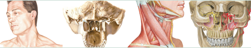
1. Head and Neck 头部与颈部
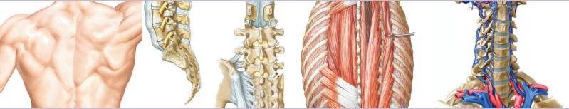
2. Back and Spinal Cord 背部与脊柱
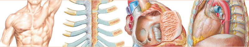
3. Thorax 胸部
4. Abdomen 腹部
5. Pelvis and Perineum 骨盆与腹膜
6. Upper Limb 上肢
7. Lower Limb 下肢
各章详细目录
Section 1. Head and Neck
-
Topographic Anatomy
-
Head and Neck: Surface Anatomy
-
-
Superficial Head and Neck
- Cutaneous Nerve of Head and Neck
- Superficial Arteries and Veins of Face and Scalp
-
Bones and Ligaments
- Skull: Anterior View
- Skull: Anteroposterior Radiograph
- Skull: Lateral View
- Skull: Lateral Radiograph
- Skull: Midsagittal Section
- Calvaria
- Cranial Base: Inferior View
- Cranial Base: Superior View
- Foramina and Canals of Cranial Base: Inferior View
- Foramina and Canals of Cranial Base: Superior View
- Skull of Newborn
- Bony Framework of Head and Neck
- Pterygoid Fossae: Posterior and Inferolateral Views
- Mandible
- Temporomandibular Joint
- Cervical Vertebrae: Atlas and Axis
- Cervical Vertebrae (continued)
- Cervical Vertebrae: Uncovertebral Joints
- External Craniocervical Ligaments
- Internal Craniocervical Ligaments
-
Superficial Face
- Facial Nerve Branches and Parotid Gland
- Muscles of Facial Expression: Lateral View
-
Neck
- Fascial Layers of Neck
- Muscles of Neck: Anterior View
- Infrahyoid and Suprahyoid Muscles
- Muscles of Neck: Lateral View
- Scalene and Prevertebral Muscles
- Superficial Veins and Cutaneous Nerves of Neck
- Nerves and Vessels of Neck
- Nerves and Vessels of Neck (continued)
- Carotid Arteries
-
Nasal Region
- Nose
- Lateral Wall of Nasal Cavity
- Lateral Wall of Nasal Cavity (continued)
- Medial Wall of Nasal Cavity (Nasal Septum)
- Nerves of Nasal Cavity
- Arteries of Nasal Cavity: Nasal Septum Turned Up
- Nerves of Nasal Cavity: Nasal Septum Turned Up
- Nose and Maxillary Sinus: Transverse Section
- Paranasal Sinuses
- Paranasal Sinuses (continued)
- Paranasal Sinuses: Changes with Age
- Salivary Glands
- Tongue and Salivary Glands: Sections
- Muscles Involved in Mastication
- Muscles Involved in Mastication (continued)
- Mandibular Nerve (V3)
- Maxillary Artery
- Ophthalmic (V1) and Maxillary (V2) Nerves
- Autonomic Innervation of Nasal Cavity
- Pterygopalatine Fossa
- Orientation of Nerves and Vessels of the Cranial Base
-
Oral Region
- Inspection of Oral Cavity
- Roof of Oral Cavity
- Floor of Oracle Cavity
- Tongue
- Tongue (continued)
- Afferent Innervation of Oral Cavity and Pharynx
- Teeth
- Teeth (continued)
-
Pharynx
- Median Section
- Muscles of Pharynx: Sagittal Section
- Pharynx: Opened Posterior View
- Muscles of Pharynx: Partial Opened Posterior View
- Fauces
- Pharyngoesophageal Junction
- Muscles of Pharynx: Lateral View
- Nerves of Oral and Pharyngeal Regions
- Arteries of Oral and Pharyngeal Regions
- Veins of Oral and Pharyngeal Regions
- Lymph Vessels and Nodes of Head and Neck
- Lymph Vessels and Nodes of Pharynx and Tongue
-
Thyroid Gland and Larynx
- Thyroid Gland: Anterior View
- Thyroid Gland and Pharynx: Posterior View
- Parathyroid Glands
- Cartilages of Larynx
- Instrisic Muscles of Larynx
- Action of Intrinsic Muscles of Larynx
- Nerves of Larynx
-
Orbit and Contents
- Eyelids
- Lacrimal Apparatus
- Fasciae of Orbit and Eyeball
- Extrinsic Eye Muscles
- Arteries and Veins of Orbit and Eyelids
- Nerves of Orbit
- Eyeball
- Anterior and Posterior Chambers of Eye
- Lens and Supporting Structures
- Intrinsic Arteries and Veins of eye
- Vascular Supply of Eye
-
Ear
- Pathway of Sound Reception
- External Ear and Tympanic Cavity
- Tympanic Cavity
- Bony and Membranous Labyrinths
-
Meninges and Brain
- Meninges and Diploic Veins
- Meningeal Arteries
- Meninges and Superficial Cerebral Veins
- Dural Venous Sinuses
- Dural Venous Sinuses (continued)
- Cerebrum: Lateral Views
- Cerebrum: Medial Views
- Cerebrum: Inferior Views
- Ventricles of Brain
- Circulation of Cerebrospinal Fluid
- Basal Nuclei (Ganglia)
- Thalamus
- Hippocampus and Fornix
- Cerebellum
- Brainstem
- Fourth Ventricle and Cerebellum
-
Cranial and Cervical Nerves
- Cranial Nerve Nuclei in Brainstem: Schema
- Crainial Nerve Nuclei in Brainstem: Schema (continued)
- Cranial Nerves (Motor and Sensory Distribution): Schema
- Olfactory Nerve (I): Schema
- Optic Nerve (II) (Visual Pathway): Schema
- Oculomotor (III), Trochlear (IV), and Abducent (VI) Nerves: Schema
- Trigeminal Nerve (V): Schema
- Facial Nerve (VII): Schema
- Vestibulocochlear Nerve (VIII): Schema
- Glossopharyngeal Nerve (IX): Schema
- Vagus Nerve (X): Schema
- Accessory Nerve (XI): Schema
- Hypoglossal Nerve (XII): Schema
- Cervical Plexus: Schema
- Autonomic Nerves in Neck
- Autonomic Nerves in Head
- Ciliary Ganglion: Schema
- Pterygopalatine and Submandibular Ganglia: Schema
- Otic Ganglion: Schema
- Taste Pathways: Schema
-
Cerebral Vasculature
- Arteries to Brain and Meninges
- Internal Carotid Artery in Petrous Part of Temporal Bone
- Arteries to Brain: Schema
- Arteries of Brain: Schema
- Arteries of Brain: Inferior Views
- Cerebral Arterial Circle (of Willis)
- Arteries of Brain: Frontal View and Section
- Arteries of Brain: Lateral and Medial Views
- Arteries of Posterior Cranial Fossa
- Veins of Posterior Cranial Fossa
- Deep Veins of Brain
- Subependymal Veins of Brain
- Hypothalamus and Hypophysis
- Arteries and Veins of Hypothalamus and Hypophysis
-
Regional Scans
- Cranial Imaging (MRV and MRA)
- Cranial Imaging (MRI)
-
Muscle Tables
Section 2. Back and Spinal Cord 背部与脊柱
-
Topographic Anatomy
- Back: Surface Anatomy
-
Bones and Ligaments
- Vertebral Column
- Thoracic Vertebrae
- Lumbar Vertebrae
- Vertebrae: Radiology
- Sacrum and Coccyx
- Vertebral Ligaments: Lumbosacral Region
- Vertebral Ligaments: Lumbar Region
-
Spinal Cord
- Spinal Cord and Ventral Rami in Situ
- Relation of Spinal Nerve Roots to Vertebrae
- Dermatomes
- Sympathetic Nervous System: Schema
- Parasympathetic Nervous System: Schema
- Spinal Membranes and Nerve Roots
- Spinal Nerve Origin: Cross Sections
- Arteries of Spinal Cord: Schema
- Arteries of Spinal Cord: Intrinsic Distribution
- Veins of Spinal Cord and Vertebral Column
- Veins of Vertebral Column: Vertebral Veins
-
Muscles and Nerves
- Muscles of Back: Superficial layers
- Muscles of Back: Intermediate Layers
- Muscles of Back: Deep Layers
- Nerves of Back
- Suboccipital Triangle
-
Cross-Sectional Anatomy
- Lumbar Region of Back: Cross Section
- Typical Thoracic Spinal Nerve: Cross Section
-
Muscle Tables
Section 3. Thorax 胸部
-
Topographic Anatomy
- Thorax: Surface Anatomy
-
Mammary Gland
- Mammary Gland
- Arteries of Mammary Gland
- Lymph Vessels and Nodes of Mammary Gland
- Lymphatic Drainage of Breast
-
Body Wall
- Bony Framework of Thorax
- Ribs and Associated Joints
- Anterior Thoracic Wall
- Anterior Thoracic Wall (continued)
- Anteriro Thoracic Wall: Internal View
- Intercostal Nerves and Arteries
- Veins of Internal Thoracic Wall
- Phrenic Nerves
- Diaphragm: Thoracic Surface
- Diaphragm: Abdominal Surface
-
Lungs
- Topographic of Lungs: Anterior View
- Topographic of Lungs: Posterior View
- Lungs in Situ: Anterior View
- Lungs: Medial Views
- Bronchopulmonary Segments
- Bronchopulmonary Segments (continued)
- Trachea and Major Bronchi
- Nomenclature of Bronchi: Schema
- Intrapulmonary Airways: Schema
- Intrapulmonary Blood Circulation: Schema
- Great Vessels of Superior Mediastinum
- Bronchial Arteries and Veins
- Lymph Vessels and Nodes of Lung
- Automonmic Nerves in Thorax
- Innervation of Tracheobronchial Tree: Schema
-
Heart
- Heart in Situ
- Heart Anterior Exposure
- Radiology and Auscultation of Heart
- Heart: Base and Diaphragmatic Surface
- Pericardial Sac
- Mediastinum: Cross Section
- Thorax: Coronal Section of Heart, Ascending Aorta
- Coronary Arteries and Cardiac Veins
- Coronary Arteries: Imaging
- Right Atrium and Ventricle
- Left Atrium and Ventricle
- Valves and Fibrous Skeleton of Heart
- Valves and Fibrous Skeleton of Heart (continued)
- Atria, Ventricles, and Interventricular Septum
- Conducting System of Heart
- Nerves of Heart
- Innervation of Heart: Schema
- Innervation of Blood Vessels: Schema
- Prenatal and Postnatal Circulation
-
Mediastinum
- Mediastinum: Right Lateral View
- Mediastinum: Left Lateral View
- Esophagus in Situ
- Topography and Constricutions of Esophagus
- Musculature of Esophagus
- Esophagogastric Junction
- Arteries of Esophagus
- Veins of Esophagus
- Nerves of Esophagus
-
Regional Scans
- Chest Scans: Axial CT Images
-
Cross-Sectional Anatomy
- Cross Section of Thorax at T3 Level
- Cross Section of Thorax at T3-4 Disc Level
- Cross Section of Thorax at T4-6 Disc Level
- Cross Section of Thorax at T7 Level
-
Muscle Table
Section 4. Abdomen 腹部
-
Topographic Anatomy
- Abdomen: Surface Anatomy
-
Body Wall
- Bony Framework of Abdomen
- Regions and Planes of Abdomen
- Anterior Abdominal Wall: Superficial Dissection
- Anterior Abdominal Wall: Intermediate Dissection
- Anterior Abdominal Wall: Deep Dissection
- Rectus Sheath: Cross Section
- Anterior Abdominal Wall: Internal View
- Posterolateral Abdominal Wall
- Arteries of Anterior Abdominal Wall
- Veins of Anterior Abdominal Wall
- Nerves of Anterior Abdominal Wall
- Thoracoabdominal Nerves
- Inguinal Region: Dissections
- Inguinal Canal and Spermatic Cord
- Femoral Sheath and Inguinal Canal
- Posterior Abdominal Wall: Internal View
- Arteries of Posterior Abdominal Wall
- Veins of Posterior Abdominal Wall
- Lymph Vessels and Nodes of Posterior Abdominal Wall
- Nerves of Posterior Abdominal Wall
-
Peritoneal Cavity
- Greater Omentum and Abdominal Viscera
- Mesenteric Relations of Intestines
- Mesenteric Relations of Intestines (continued)
- Omental Bursa: Stomach Reflected
- Omental Bursa: Cross Section
- Peritoneum of Posterior Abdominal Wall
-
Viscera (Gut)
- Stomach in Situ
- Mucosa of Stomach
- Duodenum in Situ
- Muscosa and Musculature of Duodenum
- Ileocecal Region
- Ileocecal Region (continued)
- (Vermiform) Appendix
- Mucosa and Musculature of Large Intestine
-
Viscera (Accessory Organs)
- Surface and Bed of Liver
- Liver in Situ: Vascular and Duct Systems
- Liver Structure: Schema
- Gallbladder, Extrahepatic Bile Ducts, and Pancreatic Duct
- Pancreas in Situ
- Spleen
-
Viscera Vasculature
- Arteries of Stomach, Liver, and Spleen
- Arteries of Liver, Pancreas, Duodenum, and Spleen
- Celiac Arteriogram and CT Angiogram
- Arteries of Duodenum and Head of Pancreas
- Arteries of Small Intestine
- Arteries of Large Intestine
-
Innervation
- Autonomic Nerves and Ganglia of Abdomen
- Autonomic Innervation of Stomach and Duodenum
- Autonomic Innervation of Stomach and Duodenum (continued)
- Autonomic Innervation of Stomach and Duodenum: Schema
- Autonomic Innervation of Small Intestine
- Autonomic Innervation of Large Intestine
- Autonomic Innervation of Small and Large Intestines: Schema
- Autonomic Reflex Pathways: Schema
- Intrinsic Autonomic Plexuses of Intestine: Schema
- Autonomic Innervation of Liver and Biliary Tract: Schema
- Autonomic Innervation of Pancreas: Schema
-
Kidneys and Suprarenal Glands
- Kidneys in Situ: Anterior Views
- Kidneys in Situ: Posterior Views
- Renal Artery and Vein in Situ
- Gross Structure of Kidney
- Intrarenal Arteries and Renal Segments
- Ureters in Abdomen and Pelvis
- Arteries of Ureters and Urinary Bladder
- Renal Fasciae
- Lymph Vessels and Nodes of Kidneys and Urinary Bladder
- Autonomic Nerves of Kidneys, Ureters, and Urinary Bladder
- Autonomic Innervation of Kidneys and Upper Ureters: Schema
- Autonomic Nerves of Suprarenal Glands: Dissection and Schema
- Arteries and Veins of Suprarenal Glands in Situ
-
Sectional Anatomy
- Abdominal Scans: Axial CT Images
- Abdominal Scans: Axial CT Images (continued)
- Cross Section at T10, Esophagogastric Junction
- Cross Section at T12, Inferior to Xiphoid
- Cross Section at T12-L1, Intervertebral Disc
- Cross Section at L1-2, Intervetebral Disc
- Cross Section at L3-4
-
Muscle Table
Section 5. Pelvis and Perineum 骨盆与腹膜
-
Topographic Anatomy
- Pelvis and Perineum: Surface Anatomy
-
Bones and Ligaments
- Bony Framework of Pelvis
- Radiographs of Male and Female Pelvis
- Sex Differences of Pelvis: Measurements
- Bones and Ligaments of Pelvis
- Bones and Ligaments of Pelvis (continued)
-
Pelvic Floor and Contents
- Pelvic Diaphragm: Female
- Pelvic Diaphragm: Female (continued)
- Pelvic Diaphragm: Female (continued)
- Pelvic Diaphragm: Male
- Pelvic Diaphragm: Male (continued)
- Pelvic Viscera and Perneum: Female
- Pelvic Contents: Female
- Endopelvic Fascia and Potential Spaces
- Pelvic Viscera and Perineum: Male
- Pelvic Contents: Male
-
Urinary Bladder
- Urinary Bladder: Orientation and Supports
- Female Sphincters
- Urinary Bladder: Female and Male
-
Uterus, Vagina, and Supporting Structures
- Uterine Growth
- Uterus, Vagina, and Supporting Structures
- Uterus: Fascial Ligaments
- Uterus and Adnexa
- Pelvic Ligaments
-
Perineum and External Genitalia: Female
- Female Perineum and External Genitalia (Pudendum or Vulva)
- Female Perineum (Superficial Dissection)
- Female Perineum and Deep Perineum
- Female Perneal Spaces
-
Perineum and External Genitalia: Male
- Male Perineum and External Genitalia (Superficial Dissection)
- Male Perineum and External Genitalia (Deeper Dissection)
- Penis
- Male Perineal Spaces
- Prostate and Seminal Vesicles
- Urethra
- Descent of Testis
- Scrotum and Contents
-
Homologues of Genitalia
- Homologues of External Genitalia
- Homologues of Internal Genitalia
-
Testis, Epididymis, and Ductus Deferens
- Testis, Epididymis, and Ductus Deferens
-
Rectum
- Rectum in Situ: Female and Male
- Ischio-anal Fossae
- Rectum and Anal Canal
- Anorectal Musculature
- External Anal Sphincter Muscle: Perneal Views
- Actual and Potential Perineopelvic Spaces
-
Regional Scan
- Pelvic Scans: Saggital CT Images
-
Vasculature
- Arteries of Rectum and Anal Canal: Male
- Veins of Rectum and Anal Canal: Female
- Arteries and Veins of Pelvic Organs: Female
- Arteries and Veins of Testis
- Arteries and Veins of Pelvis: Female
- Arteries and Veins of Perineum and Uterus
- Arteries and Veins of Perineum: Male
- Lymph Vessels and Nodes of Pelvis and Genitalia: Female
- Lymph Vessels and Nodes of Perineum: Female
- Lymph Vessles and Nodes of Pelvis and Genitalia: Male
-
Innervation
- Nerves of External Genitalia: Male
- Nerves of Pelvic Viscera: Male
- Nerves of Perineum: Male
- Nerves of Pelvic Viscera: Female
- Nerves of Perineum and External Genitalia: Female
- Neuropathways in Parturition
- Innervation of Female Reproductive Organs: Schema
- Innervation of Male Reproductive Organs: Schema
- Innervation of Urinary Bladder and Lower Ureter: Schema
-
Cross-Section Anatomy
- Male Pelvis: Cross Section of Bladder-Prostate Junction
- Female Pelvis: Cross Section of Vagina-Urethra
-
Muscle Tables
Section 6. Upper Limb 上肢
-
Topographic Anatomy
- Upper Limb: Surface Anatomy
-
Cutaneous Anatomy
- Dermatomes of Upper Limb
- Cutaneous Innervation of Upper Limb
- Cutaneous Nerves and Superficial Veins of Shoulder and Arm
- Cutaneous Nerves and Superficial Veins of Forearm
- Lymph Vessles and Nodes of Upper Limb
-
Shoulder and Axilla
- Clavicle and Sternoclavicular Joint
- Humerus and Scapula: Anterior Views
- Humerus and Scapula: Posterior Views
- Shoulder: Anteroposterior Radiograph
- Should (Glenohumeral Joint)
- Muscles of Shoulder
- Axilla: Posterior Wall
- Muscles of Rotator Cuff
- Pectoral, Clavipectoral, and Axillary Fasciae
- Scapulothoracic and Scapulohumeral Dissection
- Axillary Artery and Anastomoses around Scapula
- Axilla (Dissection): Anterior View
- Brachial Plexus: Schema
-
Arm
- Muscles and Arm: Anterior Views
- Muscles and Arm: Posterior Views
- Brachial Artery in Situ
- Arteries and Arm and Proximal Forearm
- Arm: Serial Cross Sections
-
Elbow and Forearm
- Bones and Elbow
- Elbow: Radiographs
- Ligaments of Elbow
- Bones of Forearm
- Individual Muscles of Forearm: Rotators of Radius
- Individual Muscles of Forearm: Extensors of Wrist and Digits
- Individual Muscles of Forearm: Flexors of Wrist
- Individual Muscles of Forearm: Flexors of Digits
- Muscles of Forearm (Superficial Layer): Posterior View
- Muscles of Forearm (Deep Layer): Posterior View
- Muscles of Forearm (Superficial Layer): Anterior View
- Muscles of Forearm (Intermediate Layer): Anterior View
- Muscles of Forearm (Deep Layer): Anterior View
- Arteries of Forearm
- Forearm: Serial Cross Sections, Anterior View
- Attachments of Muscles of Forearm: Anterior View
- Attachments of Muscles of Forearm: Posterior View
-
Wrist and Hand
- Carpal Bones
- Movements of Wrist
- Ligaments of Wrist
- Ligaments of Wrist (continued)
- Bones of Wrist and Hand
- Wrist and Hand: Radiographs
- Metacarpophalangeal and Interphalangeal Ligaments
- Wrist and Hand: Superficial Palmar Dissections
- Wrist and Hand: Deeper Palmar Dissections
- Lumbrical Muscles and Bursae, Spaces, and Sheaths: Schema
- Flexor Tendons, Arteries, and Nerves at Wrist
- Bursae, Spaces, and Tendon Sheaths of Hand
- Flexor and Extensor Tendons in Fingers
- Intrinsic Muscles of Hand
- Arteries and Nerves of Hand: Palmar Views
- Wrist and Hand: Superficial Radial Dissection
- Wrist and Hand: Superficial Dorsal Dissection
- Wrist and Hand: Deep Dorsal Dissection
- Extensor Tendons at Wrist
- Fingers
-
Neurovasculature
- Cutaneous Innervation of Wrist and Hand
- Arteries and Nerves of Upper Limb
- Nerves of Upper Limb
- Musculocutaneous Nerve
- Median Nerve
- Ulnar Nerve
- Radial Nerve in Arm and Nerves of Posterior Shoulder
- Radial Nerve in Forearm
-
Regional Scans
- Shoulder MRI, CT Scan, and Arthrogram
-
Muscle Tables
Section 7. Lower Limb 下肢
-
Topographic Anatomy
- Lower Limb: Surface Anatomy
-
Cutaneous Anatomy
- Dermatomes of Lower Limb
- Superficial Nerves and Veins of Lower Limb: Anterior View
- Superfical Nerves and Veins of Lower Limb: Posterior View
- Lymph Vessels and Nodes of Lower Limb
-
Hip and Thigh
- Hip (Coxal) Bone
- Hip Joint
- Hip Joint: Anteroposterior Radiograph
- Femur
- Bony Attachments of Muscles of Hip and Thigh: Anterior View
- Bony Attachments of Muscles of Hip and Thigh: Posterior View
- Muscles of Thigh: Anterior Views
- Muscles of Thigh: Anterior Views (continued)
- Muscles of Hip and Thigh: Lateral View
- Muscles of Hip and Thigh: Posterior Views
- Psoas and Iliacus Muscles
- Lumbosacral and Coccygeal Plexuses
- Lumbar Plexus
- Sacral and Coccygeal Plexuses
- Arteries and Nerves of Thigh: Anterior Views
- Arteries and Nerves of Thigh: Anterior Views (continued)
- Arteries and Nerves of Thigh: Posterior View
- Nerves of Hip and Buttock
- Arteries of Femoral Head and Neck
- Thigh: Serial Cross Sections
-
Knee
- Knee: Medial and Lateral Views
- Knee: Anterior Views
- Knee: Interior
- Knee: Cruciate and Collateral Ligaments
- Knee: Anteroposterior Radiograph and Posterior View
- Knee: Posterior and Sagittal Views
- Arteries of Thigh and Knee: Schema
-
Leg
- Tibia and Fibula
- Tibia and Fibula (continued)
- Attachments of Muscles of Leg
- Muscles of Leg (Superfical Dissection): Posterior View
- Muscles of Leg (Intermediate Dissection): Posterior View
- Muscles of Leg (Deep Dissection): Posterior View
- Muscles of Leg: Lateral View
- Muscles of Leg (Superficial Dissection): Anterior View
- Muscles of Leg (Deep Dissection): Anterior View
- Arteries of Knee, Leg, and Foot
- Leg: Cross Sections and Fascial Compartments
-
Ankle and Foot
- Bones of Foot
- Bones of Foot (continued)
- Calcaneus
- Ligaments and Tendons of Ankle
- Ligaments and Tendons of Foot: Plantar View
- Tendon Sheaths of Ankle
- Muscles of Dorsum of Foot: Superficial Dissection
- Dorsum of Foot: Deep Dissection
- Sole of Foot: Superficial Dissection
- Muscles of Sole of Foot: First Layer
- Muscles of Sole of Foot: Second Layer
- Muscles of Sole of Foot: Third Layer
- Interosseous Muscles and Deep Arteries of Foot
- Interosseous Muscles of Foot
-
Neurovasculature
- Femoral Nerve and Lateral Cutaneous Nerve of Thigh
- Obturator Nerve
- Sciatic Nerve and Posterior Cutaneous Nerve of Thigh
- Tibial Nerve
- Common Fibular (Peroneal) Nerve
-
Regional Scans
- Hip Radiograph, Arthrogram, and MRI
- Ankle: Radiographs
-
Muscle Tables


 7366
7366

 被折叠的 条评论
为什么被折叠?
被折叠的 条评论
为什么被折叠?
 到【灌水乐园】发言


