


传统理论认为耳鸣是由耳蜗损伤引起的,但是许多耳鸣患者表现出正常的听力图,即没有耳蜗损伤的直接迹象。最新的报告研究发现,在具有耳鸣和正常听力图的人类受试者中,听觉脑干反应显示出I波(由初级听觉神经纤维产生)的振幅显着降低,但更集中产生的V波的振幅却正常。 “隐性听力损失(hidden hearing loss)”的生理学证据表现为耳蜗神经输出减少,从而导致脑干内神经元反应幅度重新恢复正常。利用建立的计算模型,研究证明了在没有升高的听力阈值的情况下,中枢听觉系统中神经元对减少听觉神经输入的稳态反应如何引起耳鸣。
本博主以为,下面这张图,可能会成为此方面研究的经典:
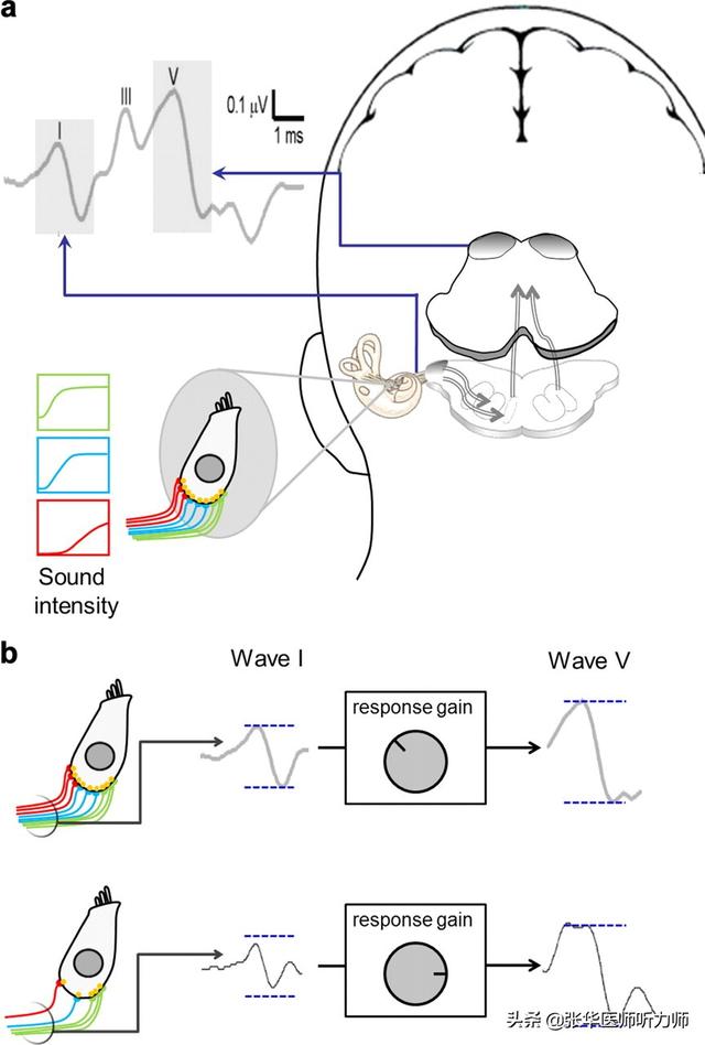
Auditory brainstem responses, hidden hearing loss, and homeostatic gain control in the auditory system. a, Illustration of the generation sites of wave I (auditory nerve) and wave V (midbrain) of the ABR, schematic depiction of an inner hair cell of the cochlea and of the AN fibers contacting it, and the rate-versus-intensity functions of the different types of auditory nerve fibers (green, low threshold fibers; blue, medium threshold fibers; red, high threshold fibers). b, Illustration of how homeostatic gain control in the auditory brainstem could normalize wave V amplitude after hidden hearing loss. In the healthy situation (top), a complete population of AN fibers gives rise to a full-sized ABR wave I, response gain in the brainstem is low, and wave V has a normal amplitude. In hidden hearing loss (bottom), a fraction of the AN fibers no longer responds to sound, leading to a reduced amplitude of ABR wave I; but through increased response gain, the amplitude of wave V has been restored to a normal size.
原文作者及出处:
Roland Schaette and David McAlpine
Journal of Neuroscience 21 September 2011, 31 (38) 13452-13457;
DOI: https://doi.org/10.1523/JNEUROSCI.2156-11.2011


 127
127


 被折叠的 条评论
为什么被折叠?
被折叠的 条评论
为什么被折叠?
 到【灌水乐园】发言


