


作者,Evil Genius
今天一大早,最新的消息来了,智联崩了。
GDP增长了5.2%,失业率屡创新高,但是可支配收入增加了,这些矛盾的背后,只有一种解释,公务员等编制类的岗位待遇上升了。
这一篇我们继续我们的多组学,单细胞、空间、外显子。参考的文章在An atlas of epithelial cell states and plasticity in lung adenocarcinoma,2024年2月发表于nature(IF 64.8)。
关于单细胞如何联合外显子的突变信息进行联合分析还需要学习学习。
其中大家如果对临床检测或者做过临检的话,对KRAS这个基因应该并不陌生,这个基因的突变导致多种癌症的发生,其中在肺腺癌中KRAS的突变,早期出现KRAS突变通常能达到临床治愈,但对于中晚期肺腺癌,出现KRAS突变一般不能治愈,KRAS突变癌细胞表现出明显的转录特征,分化减少和低水平的非整倍体。,在胰腺癌中KRAS的G12C突变有具体的靶向药可以治疗。
重复一下之前的重点,基因过量表达多发生在细胞癌变的起始阶段,基因点突变可能是细胞癌变启动阶段的一个主要事件,这一阶段的可逆性较大;对于基因表达,单细胞技术可以提供很好的帮助,对于突变,就需要借助外显子,对于免疫治疗,就需要空间、VDJ和外显子多组学的内容了。
文章研究条件
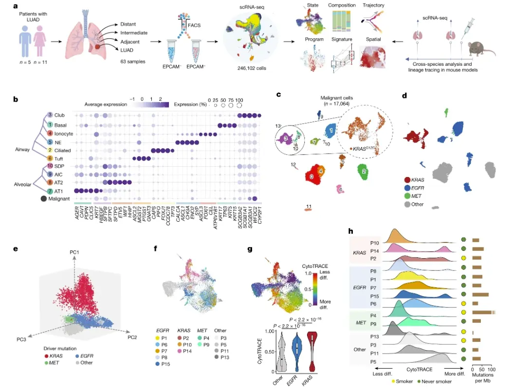
样本:来自16例早期luad和47例匹配的正常肺样本的246,102个上皮细胞(tumour-adjacent, tumour-intermediate and tumour-distant locations)。
组学:单细胞、空间(10X、DSP)、外显子、蛋白组
富集上皮细胞的基因:EPCAM
背景知识,吸烟会导致基因突变(例如KRAS)和免疫环境转变,这些变化会影响临近组织的生态环境,并且与肺恶性病变和LUAD的发展密切相关。
上皮细胞图谱
单细胞QC之后,保留246,102个上皮细胞用于分析。通过整合推断拷贝数变异(intercnv)、聚类分布、谱系特异性基因表达和携带KRASG12D体细胞突变的reads的存在等信息,将恶性细胞(n = 17,064)与非恶性正常细胞(n = 229,038)区分开来。
上皮非恶性细胞分类:alveolar、airway and a small subset of proliferative cells。其中Airway cells包括basal (KRT17+), ciliated (FOXJ1+) and club and secretory (SCGB1A1+) populations, as well as rare cell types such as ionocytes (ASCL3+), neuroendocrine cells (ASCL1+) and tuft cells (GNAT3+) 。
Alveolar cells 包括alveolar type 1 (AT1) cells (AGER1+ETV5+), AT2 cells (SFTPB+SFTPC+), SCGB1A1+SFTPC+ dual-positive cells and a cluster of alveolar intermediate cells (AICs) that was closely tucked between AT1 and AT2 clusters and shared gene expression features with both major alveolar cell types。
恶性细胞表现出标记物(markers)的低表达或不表达,总体而言,reduced lineage identity。恶性细胞形成14个clusters,主要是患者特异性的,这表明患者之间存在很强的异质性。总体而言,恶性细胞表现出高水平的非整倍体。在吸烟状况方面,没有发现任何明显的聚类模式。基于基因组图谱(WES)的注释显示,3例KRAS突变LUADs患者的恶性细胞紧密聚集在一起。相比之下,来自其他LUADs的恶性细胞表现出更分散的聚集模式。scRNA-seq分析证实,在患者特异性肿瘤clusters中存在拷贝数变异(CNV)和KRASG12D突变,而KRAS野生型LUADs (KW-LUADs)中不存在KRASG12D。
来自KM-LUADs的恶性细胞聚成一类,与EGFR突变LUADs (EM-LUADs)或MET突变LUADs (MM-LUADs)的恶性细胞明显不同。与其他LUAD相比,KM-LUADs在样本和细胞水平上都表现出更多的转录组相似性。大多数KRAS突变恶性细胞与其他细胞分开聚类,这表明KRAS突变细胞中存在不同的转录程序。与先前的报道一致,来自KM-LUADs的恶性细胞在染色体上比来自EM-LUADs的细胞更稳定。吸烟患者恶性细胞的CNV负荷明显高于不吸烟患者。恶性细胞的分化状态表现出高度的患者间异质性。也就是说,无论肿瘤突变负荷如何,KM-LUAD细胞分化程度最低,正如其最高的CytoTRACE分数所表明的那样,其次是EM-LUADs。分化状态存在肿瘤内异质性(ITH)。
An atlas of epithelial cell states and plasticity in lung adenocarcinoma
- 图例:a, Schematic overview of the experimental design and analysis workflow. Composition, composition of cell subsets; Program, transcriptional programs in malignant cells; Spatial, in situ spatial transcriptome and protein analyses; State, cellular transcriptional state. b, Proportions and average expression levels (scaled) of selected marker genes for ten normal epithelial and one malignant cell subset. NE, neuroendocrine. c, Unsupervised clustering of 17,064 malignant cells coloured by cluster identity. Top right inset shows malignant cells coloured by KRASG12D mutation status identified by scRNA-seq. d, Uniform manifold approximation and projection (UMAP) of malignant cells shown in c and coloured by driver mutations identified in each tumour sample using WES. e, Principal component analysis (PCA) plot of malignant cells coloured by driver mutations identified in each tumour sample by WES. f, UMAP plots of malignant cells coloured by patient identifier and grouped by driver mutation status. g, Top, UMAP of malignant cells by differentiation state inferred by CytoTRACE. Bottom, comparison of CytoTRACE scores between malignant cells from samples with different driver mutations. Boxes indicate the median ± interquartile range; whiskers, 1.5× the interquartile range; centre line, median. n cells in each box-and-whisker (left to right): 9,135, 5,457 and 2,472. P values were calculated using two-sided Wilcoxon rank-sum test with Benjamini–Hochberg correction. diff., differentiated. h, Per sample distribution of malignant cell CytoTRACE scores
LUAD malignant transcriptional programs
KRASG12D突变的恶性细胞分化减弱,这与KM-LUADs中肺泡分化(MP31)的丧失是一致的.来自患者P14的恶性细胞cluster表现出不同水平的CNV,其中KRASG12D细胞富集的细胞簇具有相对较晚的CNV事件(例如,染色体1p丢失,染色体8和染色体12gain),肺泡特征评分降低,结果与分化减弱一致。
AICs in LUAD
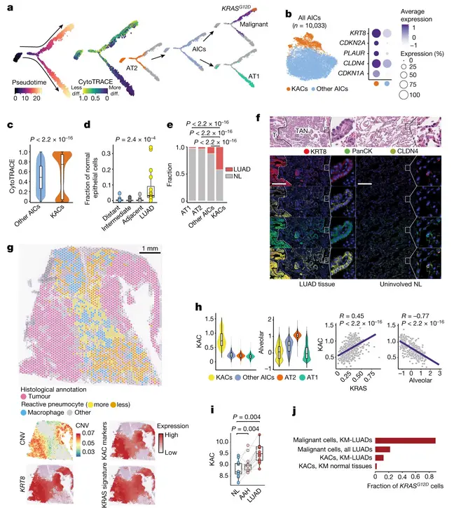
AICs在AT2 - AT1细胞发育和分化过程中处于中间位置,这一结果与暴露于急性肺损伤的无癌小鼠的中间肺泡细胞相似。LUAD组织中分化程度最低的AICs的比例高于分化程度较高的AICs。值得注意的是,AICs被推断为向恶性细胞转移,包括KRAS突变细胞,相对于EGFR突变的恶性细胞,KRAS突变细胞的发育更晚。对AICs的进一步分析发现了KRT8明显高表达的亚群。这些KACs增加了CDKN1A、CDKN2A、PLAUR和肿瘤标志物CLDN4的表达。与其他aic相比,KACs的分化程度明显较低,发育较晚。值得注意的是,KACs在轨迹推断中转化为KRAS突变的恶性细胞,而其他AIC与向AT1细胞的分化更密切相关。相对于多区NL组织,LUAD中非恶性上皮细胞中KACs的比例明显增加,且LUAD中KACs的比例明显高于AT1、AT2或其他AIC组。值得注意的是,与NL来源的KACs相比,肿瘤相关的KACs聚集在离AIC更远的地方。
为了进一步了解KACs的重要表型和特征,通过对多个肿瘤样本进行空间转录组测序(Visium spatial transcriptomics,10X)和数字空间图谱扫描(Digital spatial profiling),研究发现KACs的特征基因在mRNA水平和蛋白质水平不仅在肿瘤细胞中升高,有趣的是在肿瘤周围的临近组织中也升高,这一发现支持了文章的假设:KACs可能作为一种“前体中间态细胞”,参与早期肺腺癌的发生和发展。基于此,研究者在大量的独立数据集中进行了验证性分析。首先,利用TCGA肺腺癌数据集,发现在肺腺癌样本中KACs信号明显高于来自同一病人的配对癌旁样本。其次,在一组包含15个配对的正常肺组织,癌前病变组织,以及侵袭性肺腺癌组织的数据中,发现KACs的信号从正常肺组织到肺腺癌癌前病变组织再到早期肺腺癌组织的渐进性增强。另外,他们还发现KACs信号与病人较差的生存预后显著相关。这些结果进一步支持了研究者的假设。相关结果还显示相比不含KRAS突变的肺腺癌样本,KACs在KRAS突变型肺腺癌呈现出更高的表达水平,揭示了KAC和KRAS突变型肺腺癌之间的密切关系。
尽管与恶性细胞相比,KACs表现出较低的CNV评分,但相对于AT2、AT1和其他AIC, KACs表现出适度增加的CNV burden。KRASG12D存在于恶性细胞中,其变异等位基因频率(VAF)在KM-LUAD中为78%。KACs存在KRASG12D突变,但不包括AT2、AT1或其他AIC。KRASG12D KACs仅在KM-LUAD的组织(主要是肿瘤)中发现,因此,KRASG12D VAF(10%)在KM-LUAD的KACs中高于所有检测的LUAD的KACs(5%)或样本(3%)。在KM-LUAD患者(VAF为2%)NL样本的KACs中检测到KRASG12D突变。同时,在1例KM-LUAD患者的NL中检测到其他KRAS变体(KRASG12C),提示有潜在的区域性癌变效应。与此同时,KRASG12D型KACs的KRAS特征也比KRASWT型KACs显著增加。KRASWT KACs相对于其他AIC和其他AIC相对于AT2细胞的KRAS信号也增加。这一结果指向沿AT2-AIC-KAC频谱增加的KRAS信号。来自NL或KM-LUAD而非KW-LUAD的肿瘤的KACs与其他AIC的分化程度一致且显著低。总之,研究结果将KACs描述为与人类LUAD发病机制高度相关的中间肺泡细胞亚群,特别是KM-LUAD。
Identification and characterization of KACs in human LUAD
- 注:a, Pseudotime analysis of alveolar and malignant cells. b, Left, subclustering analysis of AICs. Right, proportions and average expression levels (scaled) of representative KAC marker genes. c, CytoTRACE score in KACs versus other AICs. n cells (left to right): 8,591 and 1,440. P value was calculated using two-sided Wilcoxon rank-sum test. d, Proportion of KACs among non-malignant epithelial cells. n samples (left to right): 16, 15, 16 and 16. P value was calculated using Kruskal–Wallis test. e, Fraction of alveolar cell subsets coloured by sample type. P values were calculated using two-sided Fisher’s exact tests with Benjamini–Hochberg correction. f, Top, haematoxylin and eosin (H&E) staining of LUAD tumour (T), TAN displaying reactive hyperplasia of AT2 cells and uninvolved NL tissue. Bottom, digital spatial profiling showing KRT8, PanCK, CLDN4, Syto13 blue nuclear stain and composite image. Magnification, ×20. Scale bar, 200 μm. Staining was repeated four times with similar results. Dashed white lines represent the margins separating tumours and TAN regions. g, ST analysis of LUAD from patient P14 showing histologically annotated H&E-stained Visium slide (left) and spatial heatmaps (right) depicting CNV score and scaled expression of KRT8, KAC markers (b) and KRAS signature. h, Expression (top) and correlation (bottom) analyses of KAC, KRAS and alveolar signatures. n = 1,440 (KACs), 8,593 (other AICs), 146,776 (AT2) and 25,561 (AT1). R, Spearman’s correlation coefficient. P values were calculated using Spearman’s correlation test. i, KAC signature expression in premalignancy cohort (15 samples each). P values were calculated using two-sided Wilcoxon signed-rank test with Benjamini–Hochberg correction. j, Fraction of KRASG12D cells in different subsets.
A KAC state is linked to mouse KM-LUAD
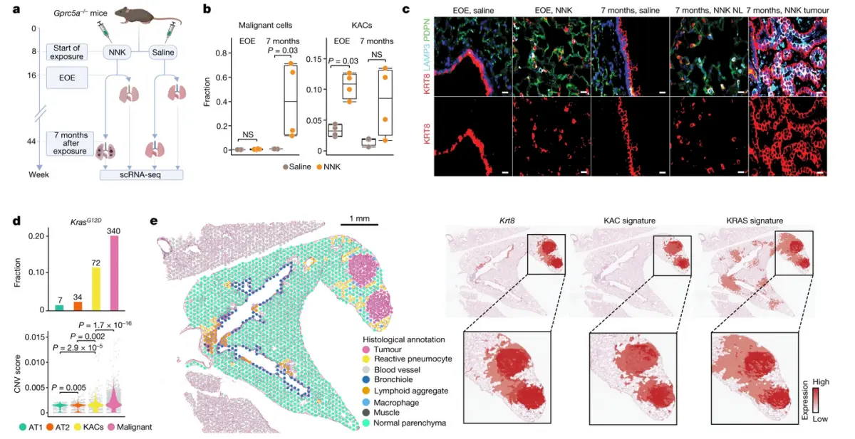
研究人员使用基因工程小鼠模型对一系列关于KACs的科学问题进行了系统探索。他们首先在一种能更准确模拟人体KRAS突变的肺腺癌发生过程的小鼠模型中进行了实验研究。该模型在烟草致癌物(Nicotine-derived nitrosamine ketone, NNK)暴露后能够适时地观察到诱导性KRAS突变型肺部肿瘤的发生。研究人员在不同时间点采集了对照和处理组样本并进行了单细胞RNA测序分析。通过对单细胞RNA测序数据的分析证实了小鼠中存在表达特征与KACs相匹配的细胞类群,研究者称之为小鼠KACs。通过对比在不同取样时间点的单细胞RNA测序数据,发现小鼠KACs的出现在KRAS突变型肿瘤出现之前。此外,研究者还对来自小鼠模型的肺癌样本进行了空间转录组测序分析。空间转录组测序数据也呈现出和人肺腺癌样本的空间转录组数据相似的特征,即小鼠肿瘤组织和肿瘤周围邻近组织中存在高水平的KACs特征基因的表达。
肺腺癌患者以及小鼠模型的单细胞RNA测序数据的拟时分析结果共同提示KACs可能来源于2型肺泡细胞。作者使用GFP标记工程小鼠,用来验证小鼠KACs是否来源于2型肺泡细胞。同时利用Gfp标记的2型肺泡细胞培养了类器官(organoids)并进行了多重免疫荧光染色分析, 实验结果显示NNK处理组的类器官样本具有更强的KACs特征基因的表达。这些结果验证了KACs的2型肺泡细胞来源并显示了NNK暴露与KACs的高度相关,揭示了KACs和肺组织损伤以及肿瘤细胞生成的密切相关。
最后,研究者构建了能够追踪KACs细胞的模型小鼠,使用谱系追踪(lineage tracing)技术回答此项研究的最终问题:KACs是否能转化成腺癌细胞。研究者使用免疫荧光染色技术分析了来自NNK暴露结束(处理组),NNK暴露后8至12周(跟进观察组)和肿瘤的样本。通过对比小鼠三组样本的免疫荧光细胞计数分析结果,研究人员发现肿瘤样本中绝大多数的细胞为2型肺泡细胞来源的,经过KACs细胞状态(Krt8+ Lamp3+)的细胞。此外,肿瘤样本中Krt8(KAC的标记基因之一)蛋白表达信号增强。谱系追踪模型小鼠的实验结果验证了2型肺泡细胞来源的KACs产生了肺腺癌细胞。
综合以上结果,这项大规模的多组学整合研究发现了KACs细胞在早期KRAS突变的肺腺癌中的重要作用,并使用转基因工程小鼠模型系统性地验证了KACs为来源于2型肺泡细胞的中间状态细胞,在2型肺泡细胞参与肺损伤修复,补充1型功能性肺泡细胞的生物学过程中,参与了Kras突变型的肺腺癌细胞的产生过程。这项研究揭示了肺腺癌发生过程和上皮细胞可塑性的重要联系,为肺腺癌的预防和早期干预提供了潜在的研究靶点。
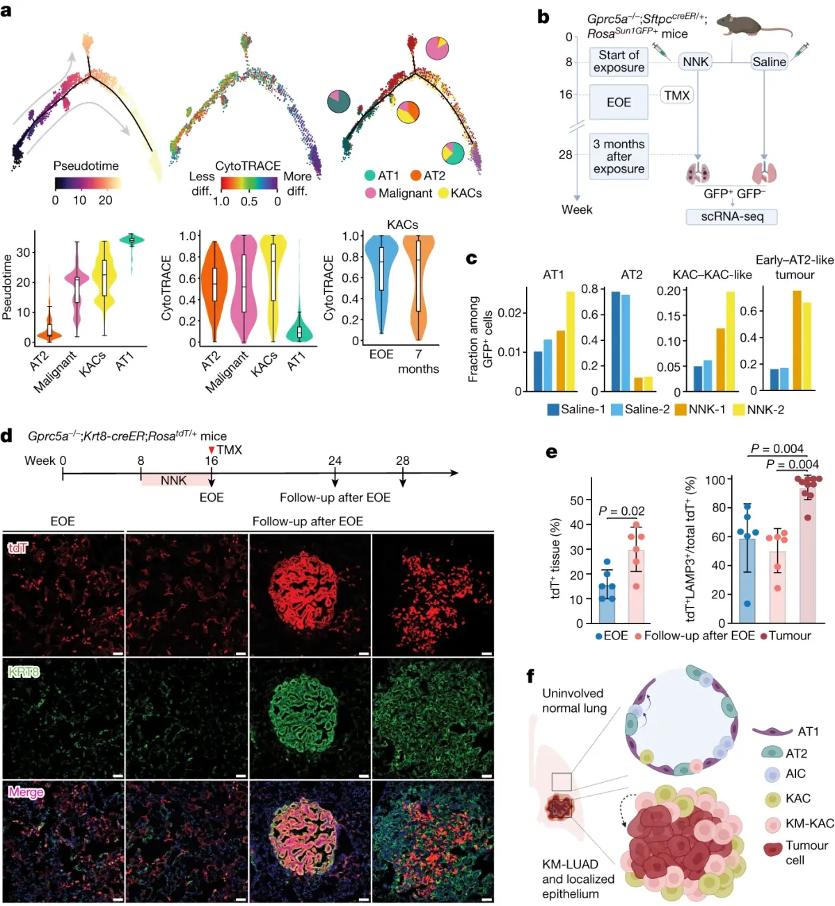
- 注:a, Trajectories of alveolar and malignant cells coloured by inferred pseudotime, cell differentiation status and cell type (top left to right). Distribution of inferred pseudotime (bottom left) and CytoTRACE (bottom middle) scores across the indicated cell subsets. Bottom right panel shows CytoTRACE score distribution in KACs at the two time points. b, Schematic overview showing analysis of Gprc5a−/− mice with reporter-labelled AT2 cells (Gprc5a−/−;SftpccreER/+;RosaSun1GFP/+). TMX, tamoxifen. c, Fractions of AT1, AT2, KACs and KAC-like cells (KAC–KAC-like) and early tumour and AT2-like tumour cells (early–AT2-like tumour) within GFP+ cells from lungs of two NNK-treated and two saline-treated mice analysed at 3 months after exposure. d, IF analysis of tdT and KRT8 expression at EOE to NNK (first column; EOE) and at 8–12 weeks following NNK (follow-up after EOE) in normal-appearing regions (second column) and tumours (last two columns) of Gprc5a−/−;Krt8-creER;RosatdT/+ mice. Tamoxifen (1 mg per dose) was delivered immediately after EOE to NNK for six continuous days. Results are representative of three biological replicates per condition. Staining was performed two times with similar results. Magnification, ×20. Scale bar, 10 μm. e, Left, percentage of lung tissue areas containing tdT+ cells. Right, percentage of tdT+LAMP3+ cells among total tdT+ cells in normal-appearing regions at different time points. Error bars show the mean ± s.d. of n biologically independent samples (left to right): 6, 6, 6, 6 and 10. P values were calculated using Mann–Whitney U-test. f, Proposed model for alveolar plasticity, whereby a subset of AICs in the intermediate AT2-to-AT1 differentiation state are KACs and, later, acquire KRASG12D mutations and are implicated in KM-LUAD development from a particular region in the lung.


 被折叠的 条评论
为什么被折叠?
被折叠的 条评论
为什么被折叠?
 到【灌水乐园】发言


