


1. 概要
DICOM标准(V3.0)已经被医疗设备生产商和医疗界广泛接受,在医疗设备中得到普及和应用。目前,越来越多的DICOM应用程序和分析软件被运用于临床医学,其中对影像的测量是临床需要的一个重要功能,例如病变的长度、肿瘤的直径等。本文依据dicom标准对Modality为XA的影像(X-Ray Angiography Image)来进行测量功能的准确性的分析。
2. DICOM标准中的X-Ray Angiographic Image信息
2.1. Composite Information Object Definitions(CIOD)
下表为X-Ray Angiographic Image在DICOM标准中规定包含的所有Modules信息。
| Modules | description | |
|---|---|---|
| This module specifies the Attributes of the Patient that describe and identify the Patient who is the subject of a Study. This Module contains Attributes of the Patient that are needed for interpretation of the Composite Instances and are common for all Studies performed on the Patient. It contains Attributes that are also included in the Patient Modules in Section C.2. | ||
| This module contains Attributes that identify a Patient as a clinical trial or research Subject. | ||
| This module specifies the Attributes that describe and identify the Study performed upon the Patient. | ||
| This module defines Attributes that provide information about the Patient at the time the Study started. | ||
| This module contains Attributes that identify a Study in the context of a clinical trial or research. | ||
| This module specifies the Attributes that identify and describe general information about the Series within a Study. | ||
| This module contains Attributes that identify a Series in the context of a clinical trial or research. | ||
| This module specifies the Attributes necessary to uniquely identify a Frame of Reference that establishes the temporal relationship of SOP Instances. A synchronized environment may be established based on a shared time of day clock, and/or on a shared trigger event or synchronization waveform channel. | ||
| This module specifies the Attributes that identify and describe the piece of equipment that produced a Series of Composite Instances. | ||
| This module specifies the Attributes that identify and describe an image within a particular Series. | ||
| This module describes the Image Pixel Module. | ||
| This module specifies the Attributes that describe the contrast /bolus used in the acquisition of the Image. | ||
| This module specifies the Attributes of a Multi-frame Cine Image. | ||
| This module specifies the Attributes of a Multi-frame pixel data Image. | ||
| This module specifies the Attributes of a Frame Pointer Module. | ||
| This module specifies the Attributes that describe mask operations for a Multi-frame image. | ||
| The Display shutter is a geometric mask consisting of one or more combined shapes that may be applied on the image for presentation purposes in order to neutralize the display of any of the pixels located outside of the shutter shape. Geometry of the shutter is specified with respect to a row and column coordinate system where the origin is the upper left hand pixel. This origin is specified by the values 1,1 for row/column. A row coordinate represents a row spacing (vertical) and a column coordinate represents a column spacing (horizontal). Up to three different shutter shapes may be used and superimposed. | ||
| This module describes the Attributes of devices or calibration objects (e.g., catheters, markers, baskets) that are associated with a Study and/or image. | ||
| This module describes the Attributes of therapies (e.g., interventions during an angiographic procedure) that are associated with a Study and/or image. | ||
| This module specifies the Attributes that identify one or more Specimens being imaged. | ||
| X-Ray Image Module. | ||
| X-Ray Acquisition Module. | ||
| An X-Ray Collimator is a device placed close to the X-Ray Source to restrict the span of the X-Ray beam. It is often made of lead shutters. This module presents in a graphical form its relationship with the Field Of View Dimensions (0018,1149). | ||
| This module contains Attributes that describe X-Ray images acquired with movement of the patient imaging table. | ||
| This module contains IOD Attributes that describe a c-arm positioner typically used in acquiring X-Ray Angiographic Images. The coordinate system used to track the positioner is defined in reference to the patient. The definition of coordinates with respect to the equipment is not supported. Furthermore, this Module does not describe the movement of the patient. | ||
| This module contains IOD Attributes that describe a DX detector. | ||
| This module contains Attributes that describe characteristics of an Overlay Plane. | ||
| This module specifies the Attributes of a Multi-frame overlay. | ||
| This module specifies the Attributes that describe the Modality LUT. | ||
| This module specifies the Attributes that describe the VOI LUT. | ||
| This module defines the Attributes that describe the hierarchical relationships of any SOP Instances referenced from other Modules within the Instance in which this Module occurs. | ||
| This module defines the Attributes that are required for proper functioning and identification of the associated SOP Instances. They do not specify any semantics about the Real-World Object represented by the IOD. | ||
| This module defines the Attributes that describe the frames extracted if the SOP Instance was created in response to a Frame-Level retrieve request. |
注:上表中M为Mandatory 、C为Conditional、U为User Option
2.2. 与测量有关的校准(Calibration )信息
从X-Ray Angiographic Image的IOD中得到与测量校准有关的数据如下表:
| tag | name | module | description |
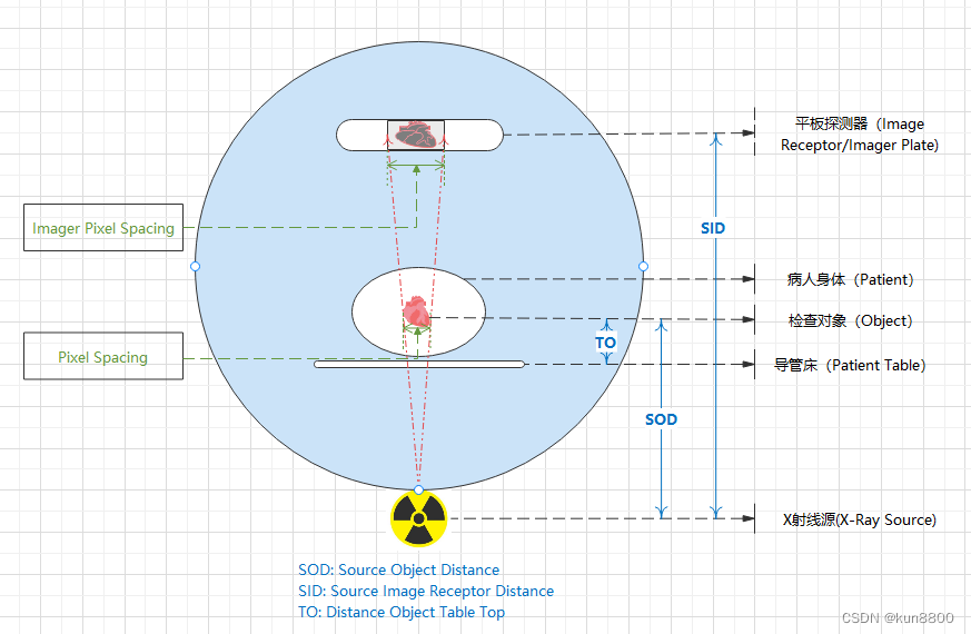
| (0018,1164) | Imager Pixel Spacing | X-Ray Acquisition | Physical distance measured at the front plane of the Image Receptor housing between the center of each pixel specified by a numeric pair - row spacing value(delimiter) column spacing value in mm. |
| (0018,1114) | Estimated Radiographic Magnification Factor | XA Positioner | Ratio of Source Image Receptor Distance (SID) over Source Object Distance (SOD). |
| (0028,0030) | Pixel Spacing | X-Ray Acquisition | Pixel Spacing (0028,0030) specifies the physical distance in the patient between the center of each pixel. |
3. 测量功能的逻辑分析
X射线源、病人和平板探测器之间的距离,平板探测器捕获的图像会被放大。这些距离越大,放大系数越大。图像上的测量值必须针对放大系数进行校准,以便根据病人的真实身体尺寸显示测量值。Image Pixel Spacing(简称IPS),是影像默认测量最有效的值,当Modality为XA的影像数据(Dataset)包含IPS以及Pixel Spacing(简称PS)时,应校验IPS的有效性,IPS 大于等于 PS视为有效。当IPS值有效及Estimated Radiographic Magnification Factor(简称ERMF)值存在当使用IPS和ERMF共同进行测量值计算。下表展示各个值相互关系:
注:IPS有效表示其标签及值都存在并且值大于等于PS的值
PS有效表示其标签及值都存在
ERMF有效表示其标签及值都存在
忽略表示不关心标签及值是否存在
| 测量准确度 | IPS | PS | ERMF | 测量结果 |
| Not calibrated: 未校准 | 无效 | 无效 | 忽略 | 无效的测量结果 |
| Calibrated: 设备校准 | 有效 | 有效 | 无效 | 依据设备校准的测量结果(更多是人为在设备上定标),使用PS进行测量计算 |
| Detector: 未校准 | 有效 | 无效 | 无效 | 比实际物理尺寸放大的测量结果,使用IPS进行测量计算 |
| Magnified: 使用ERMF校正的测量值 | 有效 | 无效 | 有效 | 依据设备校准的测量结果(没有人工参与定标,依据设备本身的参数),使用IPS和ERMF进行计算 |
| *Calibrated: 设备校准 | 无效 | 有效 | 忽略 | 依据设备校准的测量结果(更多是人为在设备上定标),但dicom影像数据存在异常,使用PS进行计算 |
| Calibrated manually: 手动校准 | 忽略 | 忽略 | 忽略 | 手动校准,准确度依据人工操作 |
当图像中存在已知物体的物理尺寸时,用户可以采取手动定标的方式来重新修正测量结果。例如冠脉手术一般会放置造影导管至主动脉的开口,这个导管的规格是已知的,那么就可以在图中测量出导管的直径后录入其实际尺寸。
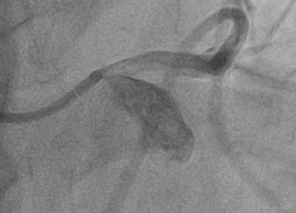
因为参照物(导管)是与检查对象在同一个空间位置(相对于射线源与探测器),那么这种校准是准确度最高的。其准确性仅受操作人员的操作影响,但是这种影响可以通过合理的操作来减小到最低,例如当校准时先将参照物放大到可以清晰标记长度的视野内,在进行校准操作。
那么对于X-Ray Angiographic Image测量的校准数据使用逻辑顺序为:
1. 手动校准
2. 使用PS
3. 使用IPS及ERMF
4. 使用IPS
5. 无效,显示像素距离
4. 结论
通过对分析X-Ray Angiographic Image类型影像的DICOM标准得知,对图像的测量结果是有不同测量准度要求的,应该按照第3节分析的结论来设计软件功能,保证用户能够明确测量的准确度信息。


 407
407


 被折叠的 条评论
为什么被折叠?
被折叠的 条评论
为什么被折叠?
 到【灌水乐园】发言


