


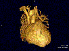
磁共振成像过程
技术 (Technology)
MRI scanners allow us, health professionals to peer into your body. Cross-sectional images of your brain, breast, knee, shoulder, or other body structure. We can see ligaments and tendons in your knee, or spy cancer in a dense breast.
M RI扫描仪使我们,卫生专业人员可以窥探您的身体。 您的大脑,乳房,膝盖,肩膀或其他身体结构的横截面图像。 我们可以看到膝盖的韧带和肌腱,或者乳房密实的间谍癌。
I had a benign brain tumor, and MRI proved invaluable in defining the target for the neurosurgeon. But, MRI can be uncomfortable. For my brain MRI, if I open my eyes, I feel like I am in a coffin. For a breast study, a woman has her arms overhead for twenty minutes or more.
我患有良性脑瘤,而MRI在确定神经外科医生的靶标方面无价之宝。 但是,MRI可能不舒服。 对于我的脑部MRI,如果睁开眼睛,我感觉就像是在棺材里。 进行乳房研究时,女人的头顶手臂会停留二十分钟或更长时间。
Before we get into how scientists are using artificial intelligence/machine learning to shorten the MRI examination time, I want to chat about magnetic resonance imaging basics.
在我们探讨科学家如何使用人工智能/机器学习来缩短MRI检查时间之前,我想谈谈磁共振成像的基础知识。

How does an MRI make images?
MRI如何制作图像?
MRI scans use extraordinarily strong magnets to make a strong magnetic field that forces protons in your cells to align with that field. When the machine then pulses a radio-frequency current through the patient, the protons become stimulated and spin out of equilibrium, as they pull against the magnetic field.
MRI扫描使用非常强的磁体产生强大的磁场,从而迫使细胞中的质子与该磁场对齐。 然后,当机器通过患者发出射频电流脉冲时,由于质子受到磁场拉动,因此质子受到刺激并失去平衡。
The technician turns off the radio-frequency field. The MRI sensors then detect the energy released as the protons realign with the magnetic field. Here’s how MRI gets its detailed images: The protons’ time to realign with the magnetic field (and the released energy) changes based on the environment and the molecules’ chemical nature. The reading radiologist can distinguish between various tissue types based on these magnetic properties.
技术人员关闭射频场。 然后,MRI传感器检测到质子与磁场重新对齐时释放的能量。 MRI如何获取其详细图像:质子与磁场(和释放的能量)重新对准的时间根据环境和分子的化学性质而变化。 阅读放射线医师可以基于这些磁性来区分各种组织类型。
To obtain an MRI image, you enter a large magnet. Hold still, or the image may be blurry. A contrast agent, often containing Gadolinium, may be given by vein before or during the MRI study — the injectable can speed up the proton realignment rate. The faster the realignment, the brighter the image appears.
要获取MRI图像,请输入一个大磁铁。 保持不动,否则图像可能模糊。 在MRI研究之前或期间,可以通过静脉注射通常含有Ga的造影剂-注射剂可加快质子重排速度。 重新对齐的速度越快,图像显示就越亮。
With an understanding of such challenges, we turn to how researchers at New York University are using AI to speed up the process. They partner with Facebook Artificial Intelligence Research to determine if speeding up the MRI scan while acquiring less data than usual can still produce images that don’t sacrifice quality.
了解了这些挑战之后,我们转向纽约大学的研究人员如何使用AI来加快这一过程。 他们与Facebook人工智能研究公司合作,确定在获取比平时少的数据的同时加快MRI扫描是否仍能产生不牺牲质量的图像。
Artificial intelligence uses machine learning. The AI analyzes the smaller volume data, creating an enhanced image for the radiologist to view. So does it work? The researchers wanted to know it doctors could tell the difference between regular MRI images and those that used artificial intelligence.
人工智能使用机器学习。 AI分析较小的体积数据,为放射线医师创建增强的图像以供查看。 这样有效吗? 研究人员想知道,医生可以分辨出常规MRI图像和使用人工智能的图像之间的区别。
The artificial intelligence-enhanced images (created using less data than is typically gathered from an MRI) did well relative to the usual MRI images.
相对于通常的MRI图像,人工智能增强的图像(使用比从MRI通常收集的数据少的数据创建的)效果很好。

The radiologists could not tell whether they were looking at accelerated or traditional pictures. The doctors could make the right diagnosis as frequently as the usual MRI approach. Of the six doctors who read the images, only one could distinguish the artificial intelligence images from the usual ones.
放射科医生无法分辨他们是在看加速图像还是传统图像。 医生可以像通常的MRI方法一样频繁地做出正确的诊断。 在读取图像的六位医生中,只有一位可以将人工智能图像与普通图像区分开。
Going forward, the researchers will take the regular MRI images and the artificial intelligence-created and see how well they predict what the surgeon sees at a knee operation. Someday, we may replace CT scans (and the ionizing radiation therapy associated with it) with fast MRI. In the breast imaging world, I look forward to Fast MRI coming to my facility within the next year.
展望未来,研究人员将拍摄常规的MRI图像和人工创建的人工智能,并观察他们对外科医生在膝盖手术中看到的图像的预测程度。 有一天,我们可能会用快速MRI代替CT扫描(以及与其相关的电离放射疗法)。 在乳腺成像领域,我期待明年快速MRI进入我的设备。
Thank you for joining me today. I’m Dr. Michael Hunter.
谢谢您今天加入我的行列。 我是Michael Hunter博士。
磁共振成像过程


 2495
2495


 被折叠的 条评论
为什么被折叠?
被折叠的 条评论
为什么被折叠?
 到【灌水乐园】发言


