


Title
题目
DIAS: A dataset and benchmark for intracranial artery segmentation in DSA sequences
DIAS:用于DSA序列中颅内动脉分割的数据集和基准
01
文献速递介绍
脑血管疾病对全球死亡率和长期残疾的贡献巨大(Vaduganathan等,2022)。近年来,得益于医学和成像技术的进步,颅内动脉(IA)疾病,如颅内动脉狭窄(ICAS)、大脑中动脉闭塞(MCAO)和颅内动脉瘤的死亡率有所下降(Hess,2018)。在这些技术中,数字减影血管造影(DSA)是一种成像方式,通过捕捉一系列图像来可视化造影剂在血管中的流动,通常持续3到15秒,采样率为每秒3到5帧(Jin等,2020)。DSA通过减去对比前的图像,有效去除了骨骼结构,从而增强了充满造影剂的血管的可见性。由于其固有的优越空间和时间分辨率,DSA在计算机断层血管造影(CTA)和磁共振血管造影(MRA)可能无法提供明确诊断的情况下,能够准确揭示病变的细节(Hess,2018)。因此,DSA被普遍认为是研究病变血管结构、解读动脉供血动态以及指导血管内介入的黄金标准(Shaban等,2022)。
Abatract
摘要
The automated segmentation of Intracranial Arteries (IA) in Digital Subtraction Angiography (DSA) playsa crucial role in the quantification of vascular morphology, significantly contributing to computer-assistedstroke research and clinical practice. Current research primarily focuses on the segmentation of single-frameDSA using proprietary datasets. However, these methods face challenges due to the inherent limitation ofsingle-frame DSA, which only partially displays vascular contrast, thereby hindering accurate vascular structurerepresentation. In this work, we introduce DIAS, a dataset specifically developed for IA segmentation in DSAsequences. We establish a comprehensive benchmark for evaluating DIAS, covering full, weak, and semisupervised segmentation methods. Specifically, we propose the vessel sequence segmentation network, in whichthe sequence feature extraction module effectively captures spatiotemporal representations of intravascularcontrast, achieving intracranial artery segmentation in 2D+Time DSA sequences. For weakly-supervised IAsegmentation, we propose a novel scribble learning-based image segmentation framework, which, under theguidance of scribble labels, employs cross pseudo-supervision and consistency regularization to improve theperformance of the segmentation network. Furthermore, we introduce the random patch-based self-trainingframework, aimed at alleviating the performance constraints encountered in IA segmentation due to thelimited availability of annotated DSA data. Our extensive experiments on the DIAS dataset demonstratethe effectiveness of these methods as potential baselines for future research and clinical applications.
在数字减影血管造影(DSA)中对颅内动脉(IA)进行自动分割在血管形态学的量化中起着关键作用,对计算机辅助的中风研究和临床实践具有重要贡献。目前的研究主要集中在使用专有数据集对单帧DSA进行分割。然而,由于单帧DSA的固有局限性,这些方法面临挑战,因为它只能部分显示血管对比,从而影响了血管结构的准确表示。在这项工作中,我们引入了DIAS,一个专门为DSA序列中的IA分割开发的数据集。我们为DIAS建立了一个全面的基准,用于评估全监督、弱监督和半监督的分割方法。具体来说,我们提出了血管序列分割网络,其中的序列特征提取模块能够有效捕捉血管内对比剂的时空表示,实现了2D+时间DSA序列中的颅内动脉分割。对于弱监督的IA分割,我们提出了一种基于涂鸦学习的新型图像分割框架,该框架在涂鸦标签的指导下,采用交叉伪监督和一致性正则化来提高分割网络的性能。此外,我们引入了基于随机补丁的自训练框架,旨在缓解由于标注的DSA数据有限而对IA分割性能造成的限制。我们在DIAS数据集上的广泛实验表明,这些方法在未来的研究和临床应用中作为潜在的基线方法是有效的。
Method
方法
Automatic IA segmentation in DSA sequences is an under-exploredand challenging task. To fully explore potential research topics, an elaborately designed benchmark for IA segmentation was proposed, aimingto provide comprehensive evaluation of the DIAS dataset using FullySupervised Segmentation (FSS), WSS, and SSS, which are increasinglydrawing attention in the medical image analysis community.
自动化颅内动脉(IA)在DSA序列中的分割是一个尚未充分探索且具有挑战性的任务。为了全面探索潜在的研究课题,我们提出了一个精心设计的IA分割基准,旨在通过全监督分割(FSS)、涂鸦学习分割(WSS)和自训练分割(SSS)对DIAS数据集进行全面评估,这些方法在医学图像分析领域正受到越来越多的关注。
Conclusion
结论
In this paper, we present the DSA-sequence IA segmentation dataset,DIAS, which provides pixel-level annotations along with two forms ofweakly supervised annotations. We establish a benchmark for evaluating IA sequence segmentation on DIAS dataset, comprising (1) the vessel sequence segmentation network, adept at capturing the spatiotemporal representations in DSA angiography; (2) the weakly-supervisedsegmentation framework integrating scribble-supervision with consistency regularization; and (3) the random patch-based self-trainingframework, strategically designed to utilize unannotated DSA datato enhance segmentation performance. Leveraging the DIAS dataset,we have conducted comprehensive experiments to validate the effectiveness of our proposed methodologies, facilitating a deeper understanding of their performance characteristics and offering valuableinsights for future research endeavors. Looking ahead, our research willcontinue, including the expansion of segmentation targets to veins andthe segmentation of arteries from complete angiographic sequences.We would like to emphasize that the DIAS dataset and its associatedbenchmark will be made publicly available, enabling researchers toreplicate our work and explore novel algorithms in this domain.
在本文中,我们介绍了用于DSA序列中颅内动脉(IA)分割的数据集DIAS,该数据集提供了像素级标注以及两种形式的弱监督标注。我们为DIAS数据集上的IA序列分割建立了一个基准,其中包括:(1) 血管序列分割网络,擅长捕捉DSA血管造影中的时空表示;(2) 弱监督分割框架,结合了涂鸦监督和一致性正则化;以及(3) 基于随机补丁的自训练框架,策略性地利用未标注的DSA数据来提高分割性能。利用DIAS数据集,我们进行了全面的实验,验证了我们提出的方法的有效性,促进了对其性能特征的深入理解,并为未来的研究工作提供了有价值的见解。展望未来,我们的研究将继续扩展分割目标,包括静脉的分割以及完整血管造影序列中的动脉分割。我们要强调的是,DIAS数据集及其相关的基准将公开提供,允许研究人员复现我们的工作并探索该领域的创新算法。
Figure
图
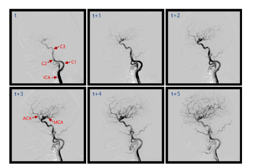
Fig. 1. A representative sample of intracranial vessels in lateral view from DSA, wheret to t+5 are consecutive frames of the arterial phase. The red arrow in the upper leftimage indicates segments C1-C3 of the intracranial internal carotid artery (ICA), whilethe lower left image represents the anterior cerebral artery (ACA) and the middlecerebral artery (MCA).
图1. DSA中颅内血管的一个代表性样本,显示了动脉相位的连续帧,从t到t+5。左上图中的红色箭头指示了颅内颈内动脉(ICA)的C1-C3段,而左下图表示前脑动脉(ACA)和中脑动脉(MCA)。
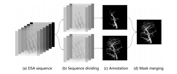
Fig. 2. Full annotated process of DIAS.
图2. DIAS的完整标注流程。
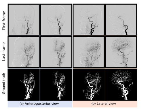
Fig. 3. Full annotated representative samples of DIAS at anteroposterior view andlateral view.
图3. DIAS在前后位和侧位的完整标注代表性样本。
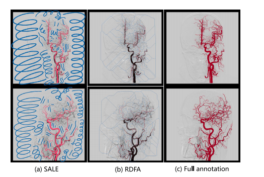
Fig. 4. Examples of two forms scribble annotations: the Scribbles Annotation with Littleclinical Experience (SALE) and Random Drawing based the Full Annotation (RDFA). Redrepresents annotated vessel pixels, blue represents annotated background pixels.
图4. 两种形式的涂鸦标注示例:基于较少临床经验的涂鸦标注(SALE)和基于完整标注的随机绘制(RDFA)。红色表示标注的血管像素,蓝色表示标注的背景像素。
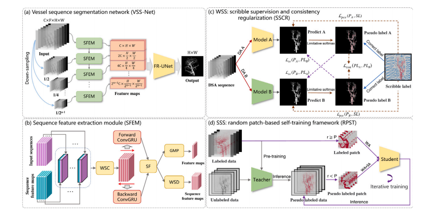
Fig. 5. Illustration of the proposed benchmark for DSA-sequence intracranial artery segmentation. (a) Vessel sequence segmentation network with (b) sequence feature extractionmodule; (c) WSS: scribble learning-based segmentation composed of scribble supervision and consistency regularization; (d) SSS: random patch-based self-training framework. (WSC:Weight Sharing Convolution, SF: Sequence Fusion, GMP: Global Max Pooling, WSD: Weight Sharing Downsampling, DA: Data augmentation, WA: Weak Augmentation, SA: StrongAugmentation).
图5. 所提出的用于DSA序列颅内动脉分割的基准示意图。(a) 血管序列分割网络,包含 (b) 序列特征提取模块;(c) WSS:基于涂鸦学习的分割,包括涂鸦监督和一致性正则化;(d) SSS:基于随机补丁的自训练框架。(WSC: 权重共享卷积, SF: 序列融合, GMP: 全局最大池化, WSD: 权重共享下采样, DA: 数据增强, WA: 弱增强, SA: 强增强)。
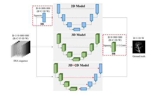
Fig. 6. Dimensionality reduction operation applied to 2D/3D models.
图6. 应用于2D/3D模型的降维操作。
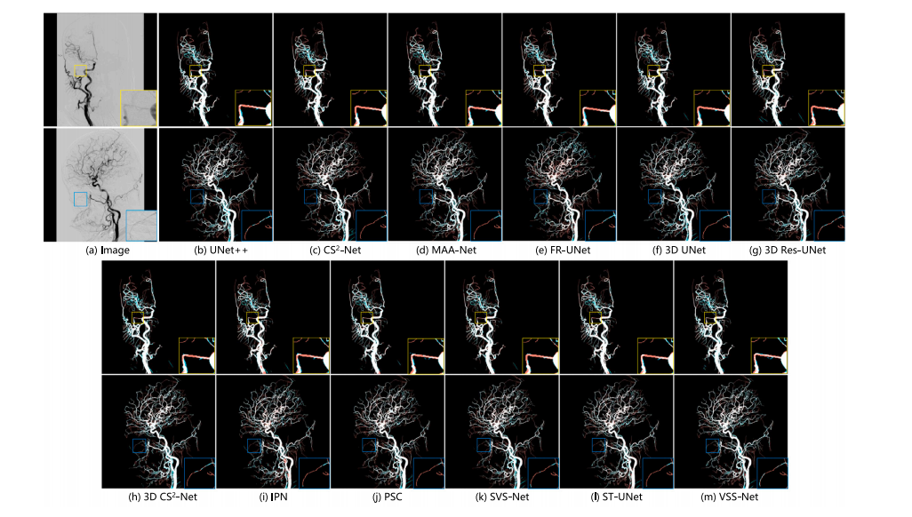
Fig. 7. Visualization of segmentation results of state-of-the-art models. The first and third rows display images of the anteroposterior view, while the second and fourth rowspresent images of the lateral view. In the segmentation maps for various methods, red pixels indicate false negatives, and green pixels signify false positives. The white and blackpixels represent correctly segmented vessels and background, respectively.
图7. 最先进模型分割结果的可视化。第一行和第三行显示了前后位图像,第二行和第四行展示了侧位图像。在各种方法的分割图中,红色像素表示假阴性,绿色像素表示假阳性。白色像素表示正确分割的血管,黑色像素表示正确分割的背景。
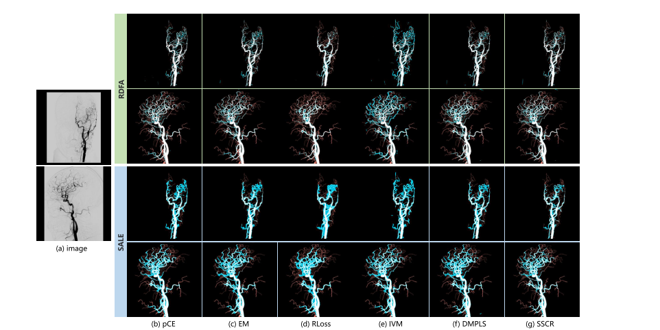
Fig. 8. Visualization of the segmentation results of scribble supervised segmentation methods trained on RDFA and SALE annotations
图8. 基于RDFA和SALE标注训练的涂鸦监督分割方法的分割结果可视化。
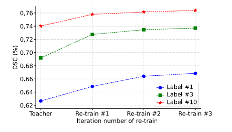
Fig. 9. Result of iterative training with different amounts of labeled data
图9. 在不同数量的标注数据下进行迭代训练的结果。
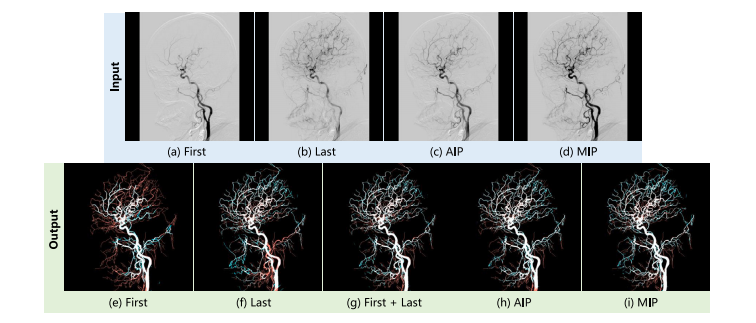
Fig. 10. The first row is the visualization of various inputs, and the second row is the corresponding segmentation result.
图10. 第一行为各种输入的可视化,第二行为相应的分割结果。
Table
表
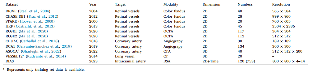
Table 1Summary of several public vessel segmentation datasets
表1 多个公开血管分割数据集的总结
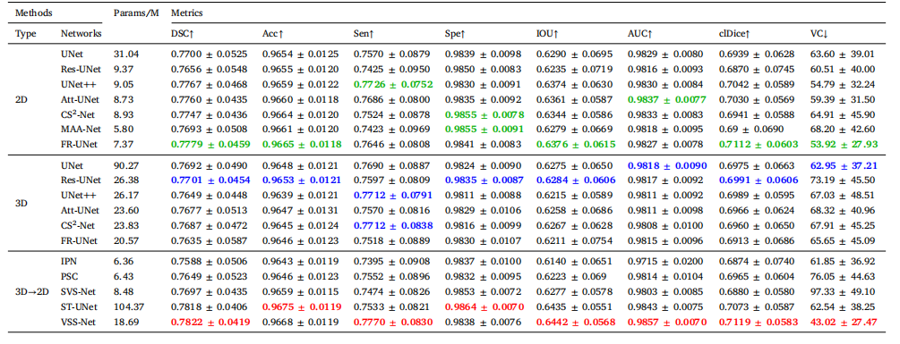
Table 2 Results of fully-supervised segmentation with state-of-the-art models. Green, blue, and red respectively represent the highest scores for each metric in 2D, 3D, and 3D→2D methods
表2 使用最先进模型进行全监督分割的结果。绿色、蓝色和红色分别代表在2D、3D和3D→2D方法中每个指标的最高分。
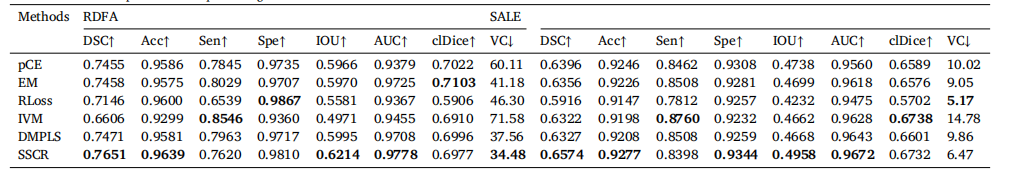
Table 3Results of Scribble-supervised IA sequence segmentation
表3 基于涂鸦监督的颅内动脉序列分割结果
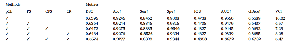
Table 4Ablation studies results of SSCR for IA sequence segmentation with SALE annotation. (pCE: partial Cross Entropy, PS: Pseudo-labeling Supervision, CPS: Cross Pseudo Supervision,CR: Consistency Regularization).
表4 基于SALE标注的SSCR颅内动脉序列分割的消融研究结果。(pCE: 部分交叉熵, PS: 伪标签监督, CPS: 交叉伪监督, CR: 一致性正则化)。
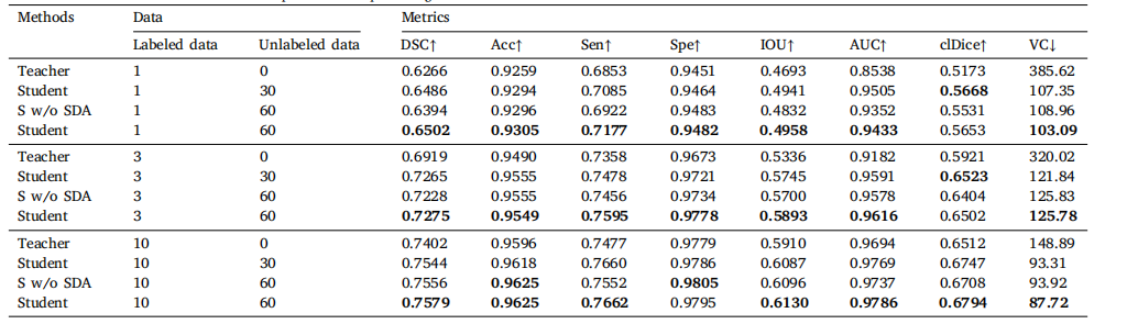
Table 5Ablation research results of RPST for semi-supervised IA sequence segmentation.
表5 半监督颅内动脉序列分割中RPST的消融研究结果。
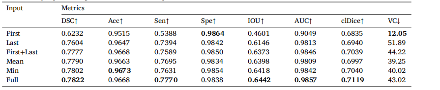
Table 6Results of fully-supervised segmentation with various input.
表6 使用各种输入进行全监督分割的结果。


 4208
4208

 被折叠的 条评论
为什么被折叠?
被折叠的 条评论
为什么被折叠?
 到【灌水乐园】发言


