


💥💥💞💞欢迎来到本博客❤️❤️💥💥
🏆博主优势:🌞🌞🌞博客内容尽量做到思维缜密,逻辑清晰,为了方便读者。
⛳️座右铭:行百里者,半于九十。
📋📋📋本文目录如下:🎁🎁🎁
目录
💥1 概述
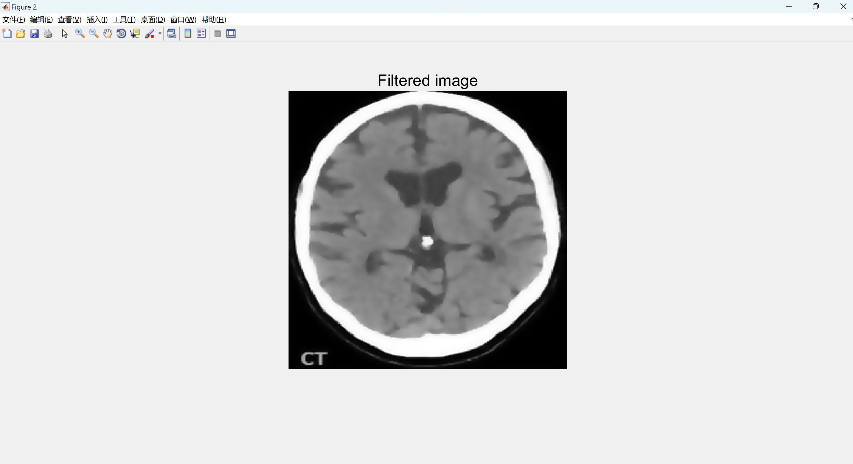
使用各向异性滤波器和图像分割技术进行MRI图像处理,可以有效地检测脑肿瘤的存在、位置、区域和边界。
摘要
脑肿瘤是一种致命的疾病,没有MRI技术,无法可靠地检测到。在这个项目中,我们尝试使用MATLAB模拟从MRI图像中检测患者大脑是否存在肿瘤。为了为MRI图像的形态学操作铺平道路,我们首先使用各向异性扩散滤波器对图像进行滤波,以降低像素之间的对比度。然后,我们调整图像大小,并手动将其转换为黑白图像,通过阈值处理来初步筛选出肿瘤可能存在的区域。
在这个半处理的图像形态学操作上,我们利用有关肿瘤大小和合理位置的信息进行进一步处理。这两个参数的最小值是根据包含肿瘤的不同MRI图像的统计平均值确定的。然后,我们使用这些参数来提供最终的检测结果。
尽管这个模拟程序在大多数情况下可以给出准确的结果,但对于过小的肿瘤或具有中空结构的肿瘤,它可能无法执行。
这个项目的更大目标是建立一个包含从不同角度拍摄的特定人类MRI图像中的肿瘤2D图像数据的数据库,并通过对这些图像进行分析来确定肿瘤的精确3D位置。为了实现这一目标,我们已经开发了2D肿瘤检测和分割方法,以提高准确性,从而使3D检测更加可靠。这是项目的主要目标。
📚2 运行结果

部分代码:
figure
imshow(erodedImage);
title('eroded image','FontSize',20);
%% subtracting eroded image from original BW image
tumorOutline=tumor;
tumorOutline(erodedImage)=0;
figure;
imshow(tumorOutline);
title('Tumor Outline','FontSize',20);
%% Inserting the outline in filtered image in green color
rgb = inp(:,:,[1 1 1]);
red = rgb(:,:,1);
red(tumorOutline)=255;
green = rgb(:,:,2);
green(tumorOutline)=0;
blue = rgb(:,:,3);
blue(tumorOutline)=0;
tumorOutlineInserted(:,:,1) = red;
tumorOutlineInserted(:,:,2) = green;
tumorOutlineInserted(:,:,3) = blue;
figure
imshow(tumorOutlineInserted);
title('Detected Tumer','FontSize',20);
%% Display Together
figure
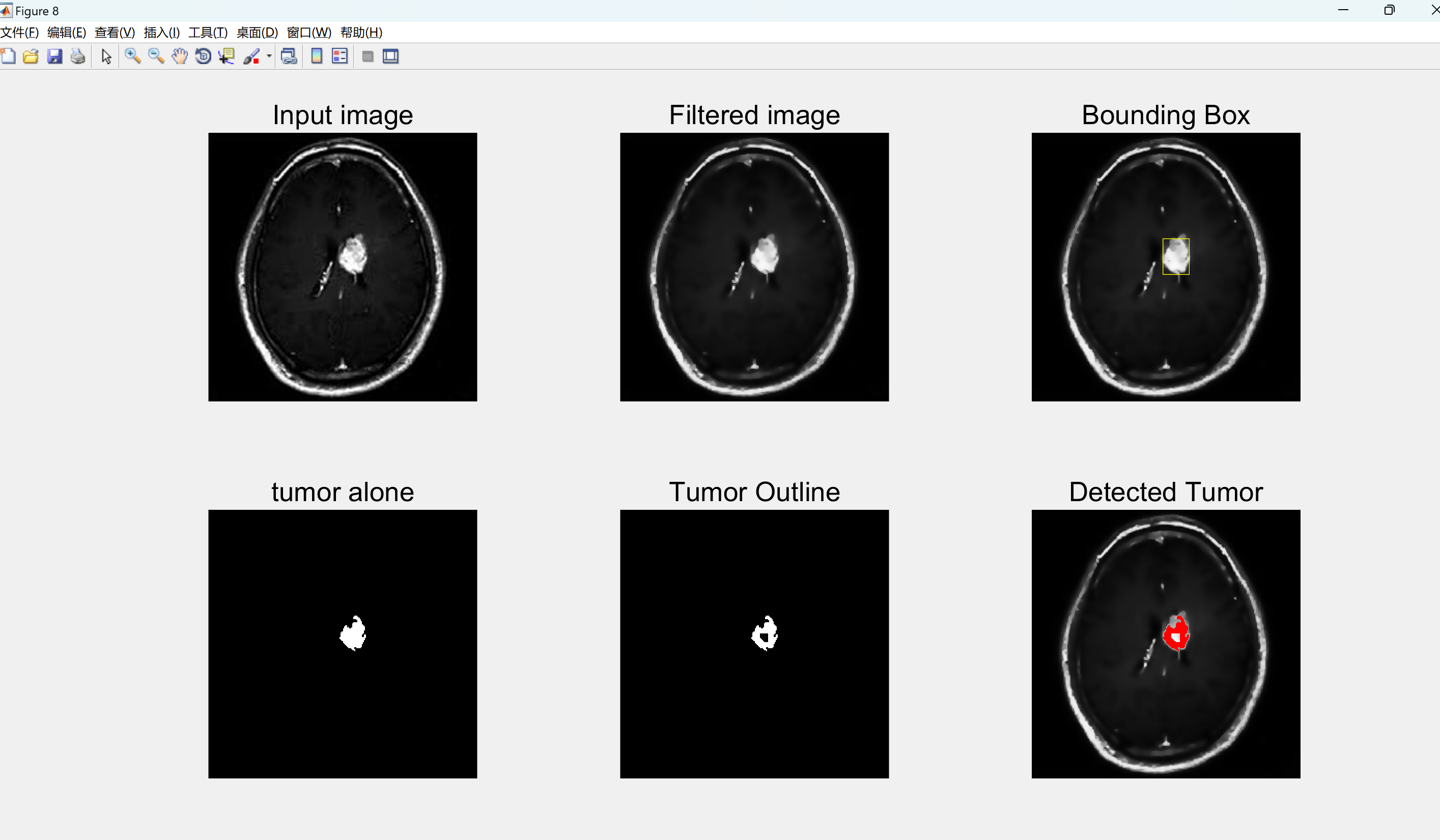
subplot(231);imshow(s);title('Input image','FontSize',20);
subplot(232);imshow(inp);title('Filtered image','FontSize',20);
subplot(233);imshow(inp);title('Bounding Box','FontSize',20);
hold on;rectangle('Position',wantedBox,'EdgeColor','y');hold off;
subplot(234);imshow(tumor);title('tumor alone','FontSize',20);
subplot(235);imshow(tumorOutline);title('Tumor Outline','FontSize',20);
subplot(236);imshow(tumorOutlineInserted);title('Detected Tumor','FontSize',20);figure
imshow(erodedImage);
title('eroded image','FontSize',20);
%% subtracting eroded image from original BW image
tumorOutline=tumor;
tumorOutline(erodedImage)=0;
figure;
imshow(tumorOutline);
title('Tumor Outline','FontSize',20);
%% Inserting the outline in filtered image in green color
rgb = inp(:,:,[1 1 1]);
red = rgb(:,:,1);
red(tumorOutline)=255;
green = rgb(:,:,2);
green(tumorOutline)=0;
blue = rgb(:,:,3);
blue(tumorOutline)=0;
tumorOutlineInserted(:,:,1) = red;
tumorOutlineInserted(:,:,2) = green;
tumorOutlineInserted(:,:,3) = blue;
figure
imshow(tumorOutlineInserted);
title('Detected Tumer','FontSize',20);
%% Display Together
figure
subplot(231);imshow(s);title('Input image','FontSize',20);
subplot(232);imshow(inp);title('Filtered image','FontSize',20);
subplot(233);imshow(inp);title('Bounding Box','FontSize',20);
hold on;rectangle('Position',wantedBox,'EdgeColor','y');hold off;
subplot(234);imshow(tumor);title('tumor alone','FontSize',20);
subplot(235);imshow(tumorOutline);title('Tumor Outline','FontSize',20);
subplot(236);imshow(tumorOutlineInserted);title('Detected Tumor','FontSize',20);
🎉3 参考文献
文章中一些内容引自网络,会注明出处或引用为参考文献,难免有未尽之处,如有不妥,请随时联系删除。
[1]周子又,刘奇,任静.基于MRI脑肿瘤的滤波方法与分割技术对比研究[J].中国医学影像学杂志, 2015, 23(7):5.DOI:10.3969/j.issn.1005-5185.2015.07.020.
[2]周子又,刘奇,任静.基于MRI脑肿瘤的滤波方法与分割技术对比研究[J].中国医学影像学杂志, 2015(007):000.
[3]曾文权,何拥军,崔晓坤.基于各向异性滤波和空间FCM的MRI图像分割方法[J].计算机应用研究, 2014, 31(1):5.DOI:10.3969/j.issn.1001-3695.2014.01.075.


 57
57


 被折叠的 条评论
为什么被折叠?
被折叠的 条评论
为什么被折叠?
 到【灌水乐园】发言


