


由于工作原因,需要做一份关于心脏的科研PPT,
正好发现了一篇2023年发表在Radiology上的文章

我们今天来接着看第三部分:结构性心脏病(Structural Heart Disease)
在之前的文章中对这篇文章的前两个章节进行了解读
#141 冠状动脉粥样硬化
-
指南文件 -
大规模临床试验 -
AI斑块分析软件 -
CAD-RADS 2.0
#142 非入侵的心脏病理
-
指南文件 -
FFR -
心肌CT灌注
我们今天来接着看第三部分:结构性心脏病(Structural Heart Disease)
1. 结构性心脏病
结构性心脏病是指解剖异常引起心脏内部组织及其发出血管的改变而造成心血管解剖、病理生理的变化。
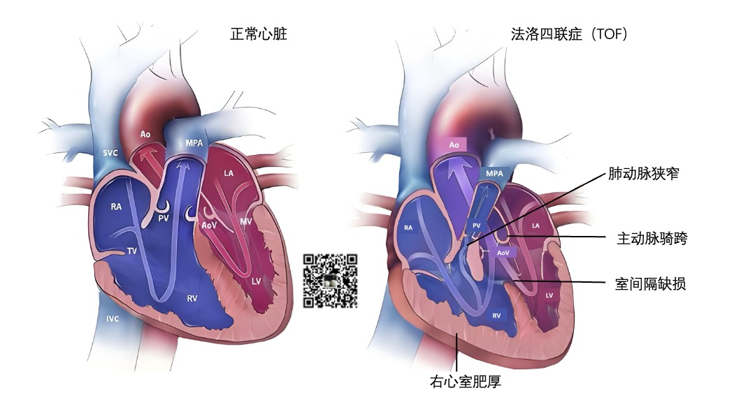
正常的心脏的解剖结构如下图:
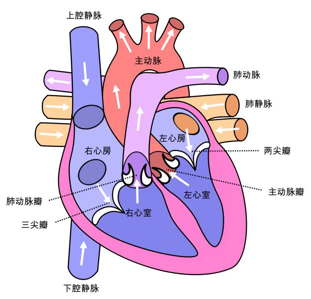
可以看到心脏有以下4个心腔(chamber),位于心脏上方的心房(ateria)较小,位于心脏下方的心室(ventricle)较大。
-
右心房(right atrium):右心房接收来自人体的低氧血液。 -
右心室(right ventricle):右心室将低氧血液泵送至肺部,使肺部充满氧气。 -
左心房(left atrium):左心房从肺部接收富含氧气的血液。 -
左心室(left ventricle):左心室将富含氧气的血液泵送至全身。
为了让血液流入或流出 4 个心腔,
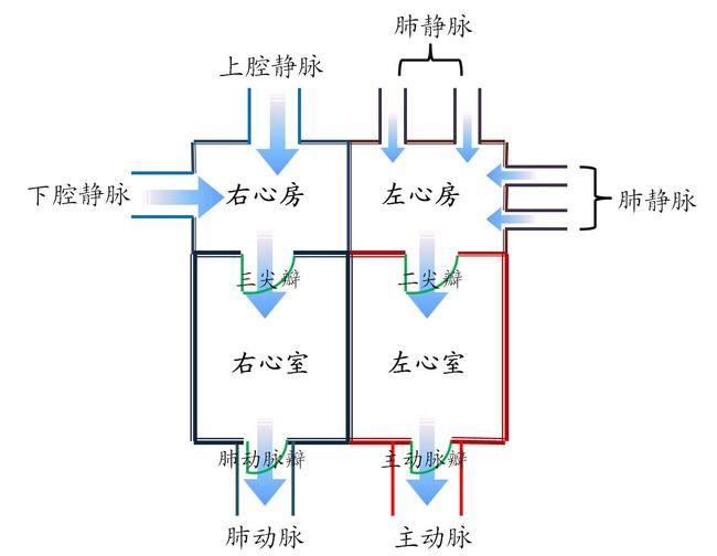
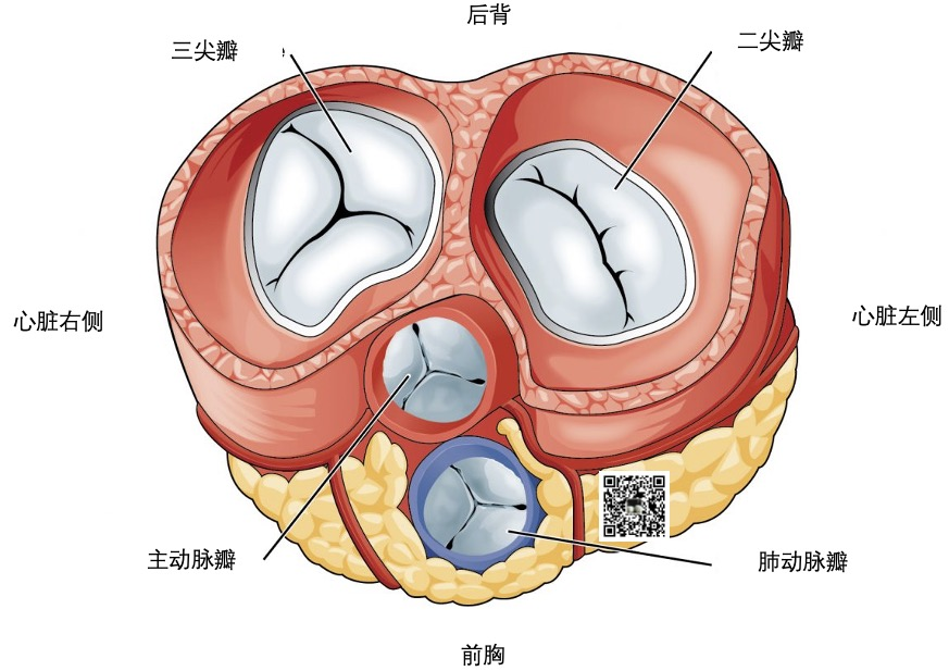
心脏有 4 个瓣膜:
-
三尖瓣(tricuspid valve):三尖瓣允许缺氧的血液从右心房流向右心室。 -
肺动脉瓣(pulmonary valve):肺动脉瓣将血液从右心室泵入肺部,使血液获得氧气。 -
二尖瓣(mitral valve):二尖瓣使富含氧气的血液从左心房流向左心室。 -
主动脉瓣(mitral valve):主动脉瓣将富含氧气的血液从左心室泵向全身。
对于结构性心脏病来说,主要包括先天性、瓣膜性心脏病、心肌病及大血管疾病等。
1. 结构性心脏病的疾病范畴
目前,葛均波院士在《结构性心脏病的定义、范畴及现状和未来》中指结构性心脏病的疾病范畴包括:
先天性心脏病
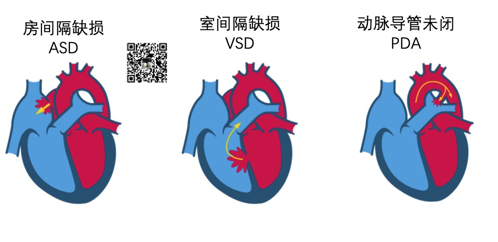
先天性心脏病主要包括室间隔缺损、房间隔缺损、动脉导管未闭,法洛氏四联症等。
房间隔缺损 (Atrial Septal Defect ,ASD)是指分隔右心房和左心房的壁上的出现破损。ASD会增加流经肺部的血液量,随着时间的推移,会损坏肺部血管。
室间隔缺损 (Ventricle Septal Defect ,VSD)是指分隔右心室和左心室出现破损。VSD使得血液可以从左心室渗入右心室,造成更多的血液被泵入肺部,迫使心脏和肺部更加努力地工作。
临床研究表明,VSD是以下疾病的高危风险因子: (1)心力衰竭 (2)中风 (3)肺动脉高压(肺部高血压) (4)心律失常(心律不齐)
动脉导管未闭(PDA)是指心脏的两条主要动脉,即主动脉和肺动脉,之间有一个连接口。通常,这个开口会在出生后不久关闭。如果没有闭合,即发生PDA。
PDA患者的主动脉中富含氧气的血液会肺动脉中缺氧的血液混合。
法洛四联症(Tetralogy of Fallot,TOF)是一种常见的先天性心脏畸形。其基本病理为室间隔缺损、肺动脉狭窄、主动脉骑跨和右心室肥厚。
心脏瓣膜病(valvular heart disease)
最常见的心脏瓣膜狭窄如下图所示:
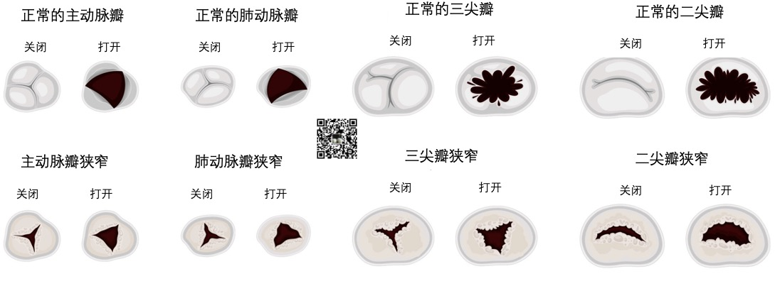
心脏瓣膜狭窄是指狭窄的瓣膜无法像正常瓣膜一样打开,使血流畅通。
随着时间的推移,这会损伤过度劳累的心肌。
最常见的主动脉瓣狭窄(Aortic Valve Stenosis)会使左心室泵血更加困难,血液无法通过瓣膜流出体外。
在肺动脉狭窄(Pulmonary Valve Stenosis)的患者中,当右心室试图将血液从肺动脉瓣泵出时,心室内的压力就会比正常情况下高得多。这时,心脏必须更加努力地将血液泵入肺部动脉。
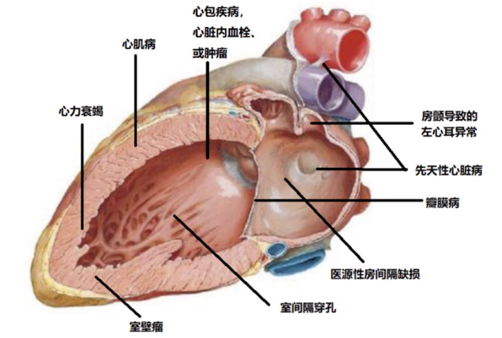
其他
-
心肌病(肥厚性心肌病、扩张型心肌病等) -
并发于其它疾病或者外源性的心脏结构异常(室间隔穿孔、室壁瘤、医源性房间隔缺损等) -
并发于其它疾病的导致心脏功能的异常并通过改变心血管结构可得到纠正的疾病或状态(如房颤导致左心耳功能异常,心力衰竭导致心脏功能的异常) -
其他更不常见的疾病:如心脏内血栓、心脏肿瘤、心包疾病等。
葛均波院士指出,由于血管外科专科和肺血管专科各有分工,因此在结构性心脏病的涉及到大血管的疾病的疾病范畴内,未包括肺血管疾病和主动脉夹层等疾病。
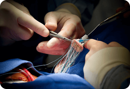
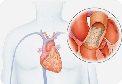
2. 结构性心脏病的治疗方式
结构性心脏病治疗包括
-
药物治疗 -
外科手术治疗
下图为手术治疗瓣膜:
-
介入治疗
目前,介入治疗已成为结构性心脏病最重要的发展方向。
就结构性心脏病介入治疗而言,主要包括以下技术:
-
先天性心脏病的经导管封堵;
-
传统的经导管瓣膜治疗术:
经皮二尖瓣球囊扩张(percutaneous balloon mitral valvuloplasty,PBMV)
经皮肺动脉瓣球囊扩张(percutaneous balloon pulmonary valvuloplasty,PBPV)
经皮主动脉瓣球囊扩张(percutaneous balloon aortic valvuloplasty,PBAV),
经导管瓣周漏封堵(Paravalvular Leak closure)等。
-
新兴的经导管瓣膜治疗术:
经导管主动脉瓣置换术(Transcatheter Aortic Valve Replacement, TAVR)
经皮肺动脉瓣置入术(Percutaneous pulmonary valve implantation,PPVI)
经导管缘对缘二尖瓣修复术(Transcatheter edge-to-edge mitral valve repair,TEER)
经导管二尖瓣置入术(Transcatheter mitral valve Replacement,TMVR)
经导管三尖瓣介入(Transcatheter Tricuspid valve implantation)。
-
经导管左心耳封堵技术(Transcatheter left atrial appendage occlusion);
-
心肌病的介入治疗:
肥厚性心肌病的酒精消融(Percutaneous transluminal septal myocardial ablation, PTSMA)
射频消融(Catheter Ablation),如
室上性心动过速导管消融术(Catheter Ablation for Supraventricular Tachycardia)
房颤导管消融术(Catheter Ablation for Atrial Fibrillation)
室性心动过速导管消融术(Catheter Ablation for Ventricular Tachycardia)
和手术混合房颤消融(Surgical Hybrid AF Ablation)等
-
心力衰竭的介入治疗: 左心室减容术
心房分流术
经导管心室辅助装置等。
3. 结构性心脏病的检查
以下检查被广泛的应用于结构性心脏病的诊断和治疗:
-
血液和尿液检查--评估包括器官功能在内的各种健康因素 -
胸部X光检查--检查心脏大小并确定有无积液 -
心电图(electrocardiogram,EGC)--记录心脏的电活动 -
超声心动图(echocardiogram,echo)--了解心脏的泵血情况
如,
心内超声心动图(intracardiac echocardiography,intracardiac echocardiography)
经食道超声心动图(transesophageal echocardiography,TEE)
经胸超声心动图(transthoracic echocardiography,TTE)等。
-
CT 成像--拍摄血流、心脏结构和运动的 X 射线图像
-
心脏核磁共振成像(Cardiac MRI)--为跳动的心脏和血管拍摄更详细的图像
-
右心导管检查(Right heart catheterisation)--检查心脏和通往肺部动脉的压力;测量心脏输出量和血氧含量。
在结构性心脏病患者的介入手术治疗流程(术前评估、术中指导和术后随访)中,综合使用上述检查方法的案例如下图所示:
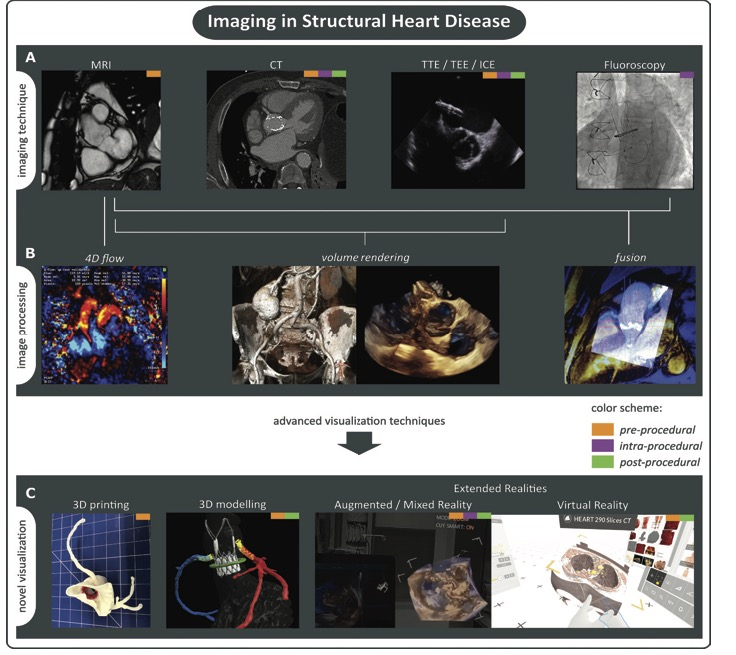
A: 目前,医生使用的主要影像检查
B: 不同的成像方法可以经过后处理增强显示或者实现不同成像技术的融合显示。
C:数据和后处理图像的临床可视化,如虚拟现实(virtual reality,VR)、增强现实(augmented reality,AR)和/或混合现实(mixed reality)、基于分割的三维建模(segmentation-based 3D modelling)和三维打印(3D printing)。
在过去的15年里,心脏CT已经从基本上与结构性干预无关,发展到在程序规划、装置尺寸和术后并发症方面发挥基础作用。
以及支持这些建议的公开证据,现在都得到了很好的证实。
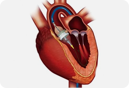
4. CT应用于TAVR
SCCT于2019年给出了用于评估心脏CT诊断TAVR的标准化循序渐进的建议共识。
| 心脏CT | 指南,共识 | 年 | 目标患者 | CT的获益 | 推荐等级 | Ref |
|---|---|---|---|---|---|---|
| 结构性心脏病 | SCCT专家共识文件 | 2019 | TAVR(transcatheter aortic valve replacement,经导管主动脉瓣膜置换术)出现严重主动脉狭窄 | 计划TAVR中的CT扫描有助于确定:1.瓣膜形态学特征;2.瓣膜的钙分布; 3.主动脉环形尺寸; 4.从主动脉环(annulus)开始的冠状动脉(coronary ostial)高度;5.主动脉和髂股动脉评估 | 强烈的专家一致性(Strong level)水平 | Ref 8 |
CT已成为术前瓣膜置换术(transcatheter aortic valve replacement ,TAVR)规划的首选成像方式。
CT上详细的解剖特征已被证明可以预测TAVR围手术期并发症:
| 风险 | 基于影像的预测因子 |
|---|---|
| (主动脉)环断裂 Annular rupture | 中度至重度LVOT(left ventricle outflow tract,左心室流出路径)钙化,CT提示面积过大超过20% |
| 冠脉闭塞/阻塞 | 冠状动脉口高度小于12mm,主动脉窦狭窄(小于30mm),结节状小叶钙化 |
| 瓣膜旁渗漏/返流 | 严重或三尖瓣小叶钙化,中度致重度LVOT钙化,环形偏心率(annular eccentricity),LVOT非管状,多层CT测量的瓣膜面积过小 |
| 房室传导异常 | 右冠状动脉尖(cusp)的钙含量较高,非冠状动脉尖钙含量低,多层CT的冠状位重建图上的膜间隔(membranous septum)长度小于8mm |
| 中风 | 主动脉弓粥样硬化程度、主动脉瓣钙化程度、左心房体积过大(新发房颤的危险因素) |
CT既可以提供主动脉根部(包括冠状动脉)的详细分析,也可以为经导管入路提供潜在的通路 (Ref 41)。
主动脉根部的详细分析
用于TAVR规划的CT数据集必须结合尽量避免瓣膜运动的主动脉环的可视化,
研究证明对于TAVR术前的主动脉瓣的测量最好是CCTA的收缩期,
对于TAVR术前的CTA扫描,扫描范围需要从锁骨下动脉开始延伸到股动脉。
在主动脉根部水平,测量的参数包括
-
主动脉环(aortic annulus) -
瓦尔萨尔瓦窦(sinuses of Valsalva) -
窦管(sino-tubular)连接
的尺寸。
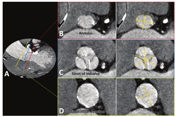
A :不同主动脉根测量的参数的横截面的位置
B:主动脉环(aortic annulus)的长径和短径(实线),周长和面积的区域(虚线)。
C:瓦尔萨尔瓦窦(sinuses of Valsalva)三条尖端到连接(cusp-to-commissure)的测量线(实线)。
D:窦管(sino-tubular)连接的长径和短径(实线),上述数值代表主动脉环到左右冠状动脉开口的距离。
独立解决方案(3mensio)和多家供应商(Vitrea、GE HealthCare)现在提供特定于TAVR的软件包,可简化上述参数的测量。
Lotus这里帮大家找到了一篇基于GE平台的TAVR软件测量的教程,感兴趣的同学可以操作一下。
-
TAVI Analysis 的功能介绍:https://mp.weixin.qq.com/s/hsQrqcrshZW2xqbpJ7tN4Q -
TAVI Analysis如何使用:https://mp.weixin.qq.com/s/HtSy023fCgoANpeHQzpZfw
如果没有半自动化的后处理工具,相关技术人员必须能够手动获得所有必要参数的测量——包括选择植入瓣膜所需的主动脉环的长轴直径、短轴直径、周长和面积。
此外,TAVR的术前分析最好还应包括瓣膜形态特征、瓣膜和(主动脉)环的钙化定量和分布、冠状动脉开口高度和手术期间使用的估计的最佳C臂投影。
TAVR入路血管评价
任何TAVR规划CT都需要对潜在的访问路径进行评估,大多数TAVR手术是通过经股动脉入路和股总动脉插管进行的。
然而,主动脉或髂动脉的扭曲可能会造成无法通过经股动脉入路实施TAVR手术,
如果是这种情况可选择的入路包括经小开胸(mini-thoracotomy)的根尖鞘置入(transapical sheath placement)、经锁骨下(trans-subclavian)、经颈动脉(transcarotid)或经下腔静脉经腹膜后交叉进入主动脉。
CT已被证明比经食管超声心动图(transesophageal echocardiography,TEE)更可靠地描绘升主动脉和主动脉弓的疾病。
因此,在最可行的TAVR入路血管评价方面,CT是一种不可或缺的成像技术。
尽管目前的研究表明,
MRI的主动脉根部和主动脉髂通路测量结果与CT具有良好的一致性,
但是CT具有高空间分辨率和广泛可用性,仍然是TAVR规划的首选成像方式。
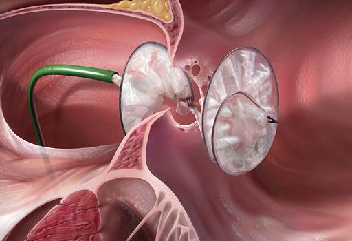
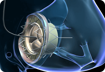
5.CT应用于二尖瓣三尖瓣的治疗
同样,心脏CT已成为较新的瓣膜置换术前不可或缺的工具,特别是对于经导管二尖瓣和三尖瓣介入治疗(Ref 42)。
例如,
经导管二尖瓣置入术存在植入后左室流出道(LVOT)阻塞和随后的心力衰竭的风险,并且是大约7%-9%的手术中可能发生的令人担忧的并发症。
当植入的装置将二尖瓣前叶移位到左心室流出道,从而使左室流出道变窄,导致所谓的新的左室流出道时,就会发生这种并发症。
如下图所示,心脏CT图像可以在术前心脏CT扫描上生成模拟的新左室流出道(Ref 43)。
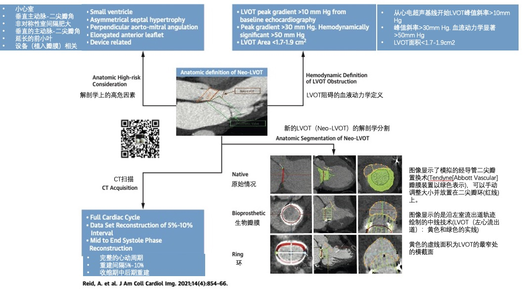
新的左心室流出道已被迅速采用为预测植入后左室流出道压差风险的关键解剖学指标,并用于瓣膜植入前的筛查。
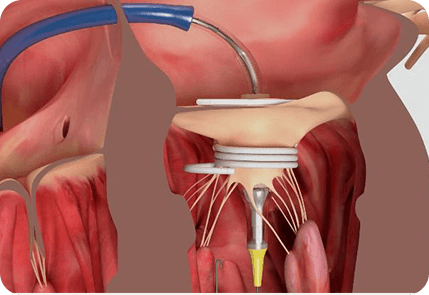
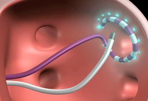
在经导管三尖瓣植入术的术前和术后评估上,CT也发挥了非常重要的作用。
下图为经导管三尖瓣植入术(EVOQUE,Edwards Lifesciences)的术前和术后CT扫描图像(Ref 44):
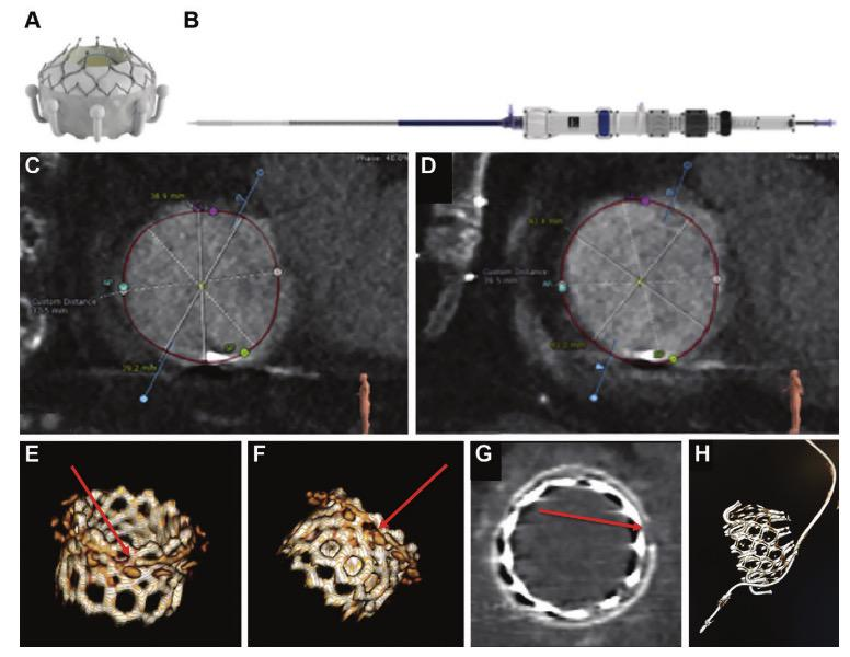
A:Evoque心脏瓣膜,包含一个自膨胀的镍钛合金框架,带有牛心包叶、环内密封裙和心室锚。
B:28-F Evoque三尖瓣系统
C:收缩期的横断面测量
D:舒张期的横断面测量
E-G: 一名因生物瓣膜植入术失败的患者接受瓣膜内SAPIEN 3瓣膜置换术,在术中需要折断生物瓣膜。
E-F:VR图显示生物瓣膜折断:生物瓣环在球囊扩张过程中被折断(箭头),以允许足够的导管瓣膜植入扩张。
G:多平面重建图显示生物瓣膜折断。
H:48-mm的EVOQUE瓣膜植入后的VR图像:术后心脏CT证实起搏器导线位于植入物外部,排除起搏器导线断裂风险。
6. CT应用于瓣膜植入术后
瓣膜植入术后的心脏 CT 还发现了瓣叶上形成的生物瓣膜血栓,即低增强瓣叶血栓(hypoattenuating valve leaflet thrombus/ hypoattenuating leaflet thickening, HALT)。
2015 年首次描述了HALT,目前对于HALT的认识仍在不断发展
如下图所示正常的TAVR叶和HALT:
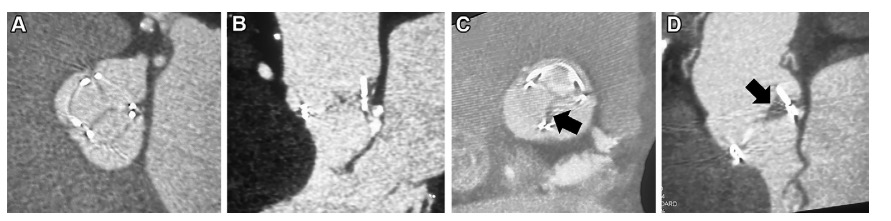
(A,B)76岁男性TAVR多平面重建显示舒张期正常TAVR叶
(C,D)84岁男性外科主动脉瓣置换术的多平面重建显示的非冠状瓣叶的异常增厚(abnormal thickening of the noncoronary leaflet)(箭头),据此可以推断出现了HALT。
这种HALT并发症可能出现在经导管和外科生物人工瓣膜上,使用抗凝治疗后即可缓解。
当HALT限制瓣叶运动时,称为影响运动的低增生( hypoattenuation affecting motion, HAM),HAM的发生可能会导致瓣膜流出道阻塞。
4D心脏 CT 使用整个心动周期的扫描数据来创建瓣叶运动的电影环路,能够有助于识别 HAM 并确定其特征,同时将其与造成手术后瓣膜坡度的其他原因(如瓣膜囊肿)区分开来。
参考文献
-
Dodd JD, Leipsic JA. Evolving Developments in Cardiac CT. Radiology. 2023;307(3):e222827. doi:10.1148/radiol.222827 IF: 12.1 Q1 -
Writing Committee Members; Gulati M, Levy PD, et al. 2021 AHA/ACC/ASE/CHEST/SAEM/SCCT/ SCMR Guideline for the Evaluation and Diagnosis of Chest Pain: Executive Summary: A Report of the American College of Cardiology/American Heart Association Joint Committee on Clinical Practice Guidelines. J Am Coll Cardiol 2021;78(22):2218–2261. -
Adamson PD, Williams MC, Dweck MR, et al. Guid- ing Therapy by Coronary CT Angiography Improves Outcomes in Patients With Stable Chest Pain. J Am Coll Cardiol 2019;74(16):2058–2070. -
DISCHARGE Trial Group; Maurovich-Horvat P, Bosserdt M, et al. CT or Invasive Coronary Angiography in Stable Chest Pain. N Engl J Med 2022;386(17):1591–1602. -
Lee SE, Sung JM, Andreini D, et al. Differential association between the pro- gression of coronary artery calcium score and coronary plaque volume pro- gression according to statins: the Progression of AtheRosclerotic PlAque DetermIned by Computed TomoGraphic Angiography Imaging (PARADIGM) study. Eur Heart J Cardiovasc Imaging 2019;20(11):1307–1314. -
Han D, Chen B, Gransar H, et al. Prognostic significance of plaque location in non-obstructive coronary artery disease: from the CONFIRM registry. Eur Heart J Cardiovasc Imaging 2022;23(9):1240–1247. -
Patel AR, Bamberg F, Branch K, et al. Society of cardiovascular computed tomography expert consensus document on myocardial computed tomography perfusion imaging. J Cardiovasc Comput Tomogr 2020;14(1):87–100. -
Blanke P, Weir-McCall JR, Achenbach S, et al. Computed tomography imag- ing in the context of transcatheter aortic valve implantation (TAVI) / transcatheter aortic valve replacement (TAVR): An expert consensus document of the Society of Cardiovascular Computed Tomography. J Cardiovasc Comput Tomogr 2019;13(1):1–20. -
Boogers MJ, Broersen A, van Velzen JE, de Graaf FR, El-Naggar HM, Kitslaar PH, et al. Automated quantification of coronary plaque with computed tomography: comparison with intravascular ultrasound using a dedicated registration algorithm for fusion-based quantification. Eur Heart J 2012;33:1007-1016 -
de Graaf MA, Broersen A, Kitslaar PH, Roos CJ, Dijkstra J, Lelieveldt BP, et al. Automatic quantification and characterization of coronary atherosclerosis with computed tomography coronary angiography: cross-correlation with intravascular ultrasound virtual histology. Int J Cardiovasc Imaging 2013;29:1177-1190 -
Fujimoto S, Kondo T, Kodama T, Fujisawa Y, Groarke J, Kumamaru KK, et al. A novel method for non-invasive plaque morphology analysis by coronary computed tomography angiography. Int J Cardiovasc Imaging 2014;30:1373-1382 -
Voros S, Rinehart S, Qian Z, Vazquez G, Anderson H, Murrieta L, et al. Prospective validation of standardized, 3-dimensional, quantitative coronary computed tomographic plaque measurements using radiofrequency backscatter intravascular ultrasound as reference standard in intermediate coronary arterial lesions: results from the ATLANTA (assessment of tissue characteristics, lesion morphology, and hemodynamics by angiography with fractional flow reserve, intravascular ultrasound and virtual histology, and noninvasive computed tomography in atherosclerotic plaques) I study. JACC Cardiovasc Interv 2011;4:198-208 -
Choi AD, Marques H, Kumar V, Griffin WF, Rahban H, Karlsberg RP, et al. CT evaluation by artificial intelligence for atherosclerosis, stenosis and vascular morphology (CLARIFY): a multi-center, international study. J Cardiovasc Comput Tomogr 2021;15:470-476 -
Sheahan M, Ma X, Paik D, Obuchowski NA, St Pierre S, Newman WP 3rd, et al. Atherosclerotic plaque tissue: noninvasive quantitative assessment of characteristics with software-aided measurements from conventional CT angiography. Radiology 2018;286:622-631 -
Dey D, Schepis T, Marwan M, Slomka PJ, Berman DS, Achenbach S. Automated three-dimensional quantification of noncalcified coronary plaque from coronary CT angiography: comparison with intravascular US. Radiology 2010;257:516-522 -
Tzimas G, Gulsin GS, Everett RJ, Akodad M, Meier D, Sewnarain K, et al. Age- and sex-specific nomographic CT quantitative plaque data from a large international cohort. JACC Cardiovasc Imaging 2024;17:165-175 -
Tzimas G. Nomographic CT quantitative plaque data from a large international population. Society of Cardiovascular Computed Tomography Annual Meeting, Las Vegas, 2022. https://cdn.ymaws.com/scct.org/resource/resmgr/ scct_2022_printed_program_AB.pdf. -
Cury RC, Leipsic J, Abbara S, et al. CAD-RADSTM 2.0 - 2022 Coronary Artery Disease-Reporting and Data System: An Expert Consensus Document of the Society of Cardiovascular Computed Tomography (SCCT), the American College of Cardiology (ACC), the American College of Radiology (ACR), and the North America Society of Cardiovascular Imaging (NASCI). J Cardiovasc Comput Tomogr 2022;16(6):536–557. -
Curzen N, Nicholas Z, Stuart B, et al. Fractional flow reserve derived from computed tomography coronary angiography in the assessment and management of stable chest pain: the FORECAST randomized trial. Eur Heart J. 2021;42(37):3844-3852. doi:10.1093/eurheartj/ehab444IF: 37.6 Q1 -
Nanna MG, Vemulapalli S, Fordyce CB, et al. The prospective randomized trial of the optimal evaluation of cardiac symptoms and revascularization: Rationale and design of the PRECISE trial. Am Heart J. 2022;245:136-148. doi:10.1016/j.ahj.2021.12.004IF: 3.7 Q1 -
Bech GJW, De Bruyne B, Pijls NH, et al. Fractional flow reserve to determine the appropriateness of angioplasty in moderate coronary stenosis: a randomized trial. Circulation 2001; 103:2928–2934 -
Tonino PAL, De Bruyne B, Pijls NHJ, et al.; FAME Study Investigators. Fractional flow reserve versus angiography for guiding percutaneous coronary intervention. N Engl J Med 2009; 360:213–224 -
De Bruyne B, Pijls NHJ, Kalesan B, et al.; FAME 2 Trial Investigators. Fractional flow reserve-guided PCI versus medical therapy in stable coronary disease. N Engl J Med 2012; 367:991–1001 -
Takx RA, Blomberg BA, El Aidi H, Habets J, et al. Diagnostic accuracy of stress myocardial perfusion imaging compared to invasive coronary angiography with fractional flow reserve met- analysis. Circ Cardiovasc Imaging 2015;8:1–7. -
Koo BK, Erglis A, Doh JH, et al. Diagnosis of ischemia-causing coronary stenoses by noninvasive fractional flow reserve computed from coronary computed tomographic angiograms. Results from the prospective multi-center DISCOVER-FLOW (Diagnosis of Ischemia-Causing Stenoses Obtained Via Noninvasive Fractional Flow Reserve) study. J Am Coll Cardiol 2011; 58:1989–1997 -
Min JK, Leipsic J, Pencina MJ, et al. Diagnostic accuracy of fractional flow reserve from anatomic CT angiography. JAMA 2012; 308:1237–1245 -
Nørgaard BL, Leipsic J, Gaur S, et al.; NXT Trial Study Group. Diagnostic performance of noninvasive fractional flow reserve derived from coronary computed tomography angiography in suspected coronary artery disease: the NXT trial (analysis of coronary blood flow using CT angiography: next steps). J Am Coll Cardiol 2014; 63:1145–1155 -
Griffin WF, Choi AD, Riess JS, et al. AI Evaluation of Stenosis on Coronary CTA, Comparison With Quantitative Coronary Angiography and Fractional Flow Reserve: A CREDENCE Trial Substudy. JACC Cardiovasc Imaging. 2023;16(2):193–205. doi: 10.1016/j.jcmg.2021.10.020 IF: 12.8 Q1 . -
Madsen KT, Nørgaard BL, Øvrehus KA, et al. Prognostic Value of Coronary CT Angiography-derived Fractional Flow Reserve on 3-year Outcomes in Patients with Stable Angina. Radiology. 2023;308(3):e230524. doi: 10.1148/radiol.230524 IF: 12.1 Q1 . -
Cherukuri L, Birudaraju D, Kinninger A, et al. Use of Advanced CT Technology to Evaluate Left Atrial Indices in Patients with a High Heart Rate or with Heart Rate Variability: The Converge Registry. J Nucl Med Technol. 2021;49(1):65–69. doi: 10.2967/jnmt.120.253781 . -
Sand NPR, Veien KT, Nielsen SS, et al. Prospective comparison of FFR de- rived from coronary CT angiography with SPECT perfusion imaging in stable coronary artery disease: the ReASSESS study. JACC Cardiovasc Imaging 2018; 11:1640–1650 -
Artzner C, Daubert M, Ehieli W, et al. Impact of computed tomography (CT)-derived fractional flow reserve on reader confidence for interpreta- tion of coronary CT angiography. Eur J Radiol 2018; 108:242–248 -
Curzen NP, Nolan J, Zaman AG, Nørgaard BL, Rajani R. Does the routine availability of CT-derived FFR influence management of patients with stable chest pain compared to CT angiography alone? The CT-FFR RIPCORD study. JACC Cardiovasc Imaging 2016; 9:1188–1194 -
Douglas PS, Pontone G, Hlatky MA, et al.; PLATFORM Investigators. Clinical outcomes of fractional flow reserve by computed tomographic angiography-guided diagnostic strategies vs. usual care in patients with suspected coronary artery disease: the prospective longitudinal trial of FFR(CT)— outcome and resource impacts study. Eur Heart J 2015; 36:3359–3367 -
Andreini D, Modolo R, Katagiri Y, et al.; SYNTAX III REVOLUTION Investiga- tors. Impact of fractional flow reserve derived from coronary computed tomography angiography on heart team treatment decision-making in patients with multivessel coronary artery disease: insights from the SYNTAX III REVOLUTION trial. Circ Cardiovasc Interv 2019; 12:e007607 -
Dewey M, Siebes M, Kachelrieß M, et al. Clinical quantitative cardiac imaging for the assessment of myocardial ischaemia. Nat Rev Cardiol 2020;17(7):427–450. -
Nous FMA, Geisler T, Kruk MBP, et al. Dynamic Myocardial Perfusion CT for the Detection of Hemodynamically Significant Coronary Artery Disease. JACC Cardiovasc Imaging 2022;15(1):75–87. -
Andreini D, Mushtaq S, Pontone G, et al. CT Perfusion Versus Coronary CT Angiography in Patients With Suspected In-Stent Restenosis or CAD Progres- sion. JACC Cardiovasc Imaging 2020;13(3):732–742. -
Narula J, Chandrashekhar Y, Ahmadi A, et al. SCCT 2021 Expert Consensus Document on Coronary Computed Tomographic Angiography: A Report of the Society of Cardiovascular Computed Tomography. J Cardiovasc Comput Tomogr 2021;15(3):192–217. -
中华医学会放射学分会心胸学组, 国家心血管病专业质控中心心血管影像质控专家工作组 . 动态 CT 心肌灌注成像技术操作与图像分析中国专家共识[J]. 中华放射学杂志, 2022, 56(12): 1289-1299. DOI: 10.3760/cma.j.cn112149-20220308-00213 . -
Rudzinski PN, Leipsic JA, Schoepf UJ, et al. CT in Transcatheter-delivered Treatment of Valvular Heart Disease. Radiology. 2022;304(1):4-17. doi:10.1148/radiol.210567 -
Fam NP, von Bardeleben RS, Hensey M, et al. Transfemoral Transcatheter Tricuspid Valve Replacement With the EVOQUE System: A Multicenter, Observational, First-in-Human Experience. JACC Cardiovasc Interv 2021;14(5):501–511. -
Reid A , Ben ZekryS , Turaga M, et al . Neo-LVOT and Transcatheter Mitral Valve Replacement: Expert Recommendations. JACC Cardiovasc Imaging 2021;14(4):854–866. -
Hensey M, Alenezi AR, Murdoch DJ, et al. Transcatheter Tricuspid Valve-in- Valve Replacement With Subsequent Bioprosthetic Valve Fracture to Opti- mize Hemodynamic Function. JACC Cardiovasc Interv 2018;11(21):2226– 2227.
延伸阅读:
#141 冠状动脉粥样硬化
-
指南文件 -
大规模临床试验 -
AI斑块分析软件 -
CAD-RADS 2.0
#142 非入侵的心脏病理
-
指南文件 -
FFR -
心肌CT灌注
本文由 mdnice 多平台发布


 1700
1700

 被折叠的 条评论
为什么被折叠?
被折叠的 条评论
为什么被折叠?
 到【灌水乐园】发言


