


👨🎓个人主页:研学社的博客
💥💥💞💞欢迎来到本博客❤️❤️💥💥
🏆博主优势:🌞🌞🌞博客内容尽量做到思维缜密,逻辑清晰,为了方便读者。
⛳️座右铭:行百里者,半于九十。
📋📋📋本文目录如下:🎁🎁🎁
目录
💥1 概述
文献来源:

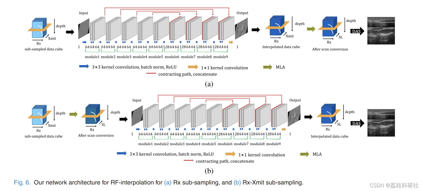
在便携式、3-D和超快速超声成像系统中,由于接收器(Rx)或发射(Xmit)事件子采样,对从有限数量的射频(RF)测量中重建高质量图像的需求日益增加。然而,由于RF子采样存在旁瓣伪影,标准波束形成器通常会产生对比度较低的模糊图像,这不适合诊断目的。现有的压缩传感方法通常需要硬件更改或计算昂贵的算法,但它们的质量改进有限。为了解决这个问题,在本文中,我们提出了一种新颖的深度学习方法,该方法利用Rx-Xmit平面中的冗余直接插值丢失的RF数据。我们使用来自多线采集B模系统的子采样RF数据进行了广泛的实验结果,证实所提方法可以在不牺牲图像质量的情况下有效降低数据速率。
原文摘要:
Abstract:
In portable, 3-D, and ultra-fast ultrasound imaging systems, there is an increasing demand for the reconstruction of high-quality images from a limited number of radio-frequency (RF) measurements due to receiver (Rx) or transmit (Xmit) event sub-sampling. However, due to the presence of side lobe artifacts from RF sub-sampling, the standard beamformer often produces blurry images with less contrast, which are unsuitable for diagnostic purposes. Existing compressed sensing approaches often require either hardware changes or computationally expensive algorithms, but their quality improvements are limited. To address this problem, in this paper, we propose a novel deep learning approach that directly interpolates the missing RF data by utilizing redundancy in the Rx-Xmit plane. Our extensive experimental results using sub-sampled RF data from a multi-line acquisition B-mode system confirm that the proposed method can effectively reduce the data rate without sacrificing the image quality.
由于超声(US)成像具有出色的时间分辨率、合理的图像质量和最小的侵入性,已成为诊断心脏、肝脏等许多疾病的黄金标准。因此,已经有许多研究工作将美国成像扩展到新的应用,例如紧急护理中的便携式成像[2],3-D成像[3],超快速成像[4],[5]等。
为了在US成像中获得更好的空间分辨率,美国传感器的Rx部分应使用高速模数转换器(ADC),这消耗大量功率。因此,在便携式美国系统中,使用少量具有减小光圈尺寸的Rx元件来降低功耗,这通常会导致图像质量下降。另一方面,为了实现更高的帧速率,应减少传输事件的数量,因为传输事件的持续时间由声速决定。这反过来又会导致伪影下采样。
为了解决这些问题,已经研究了压缩传感(CS)方法[6]-[10]。然而,美国特有的特性通常会降低CS方法的性能。例如,由于超声波散射的波性质,通常很难对传感矩阵进行精确建模。此外,美国图像包含特征斑点,这使得它们在任何基础上几乎不稀疏。瓦格纳等人没有使用波散射物理学。[8] 将扫描线轮廓建模为具有有限创新率 (FRI) 的信号 [11],并提出了一种专门设计的硬件架构,可实现高分辨率扫描线重建 [8]。然而,这种方法的缺点之一是它不能用于传统的B模式成像系统。
📚2 运行结果
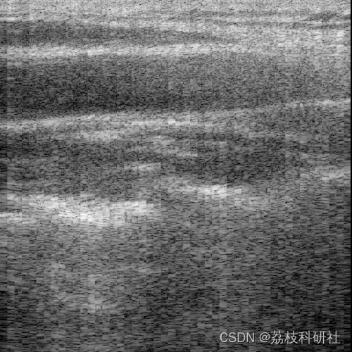
部分代码:
clc
clear all
close all
dataFile = ['data\cnn_sparse_view_init_multi_normal_dsr2_input64\DNN4x1_TestVal.mat'];
load(dataFile);
rec = images.data; % change this for images.label for label and for reconstructed image use rec
[nNumCh,ScanlineNum,Numframes,AlignedSampleNum]=size(rec);
Reconstruction = permute(2048*rec,[1 4 2 3]);
%%
Rx_F_num = 1;
Offset = 50;
fs = 40e6;
c = 1540;
% N_ele = double(System.Transducer.elementCnt);
N_ele = 192;
% pitch = double(System.Transducer.elementPitchCm) * 1e-2; % cm => m
pitch = 0.0200 * 1e-2; % cm => m
AlignedSample = double(AlignedSampleNum);
SampleNum = 2469;
DepSample = double(SampleNum);
nNumCh = 64;
N_ch = nNumCh;
nHalfNumCh = nNumCh/2;
sam_st_2nd = AlignedSample*nHalfNumCh;
data_total = AlignedSample;
data_total1 = DepSample;
pixel_d = c/fs/2;
%%
scan_view_size = pitch*N_ele; % Lateral View Size
N_sc = double(ScanlineNum);
% N_sc = ((scan_view_size/pitch)) ; % Scanline number
sc_d = scan_view_size/(N_sc); % Scanline distance
st_sc_x = - scan_view_size/2+sc_d/2 ; % Start Scanline position
st_sam = round(0.001/pixel_d);
ed_sam = data_total1;
%% DC cancle filter
f = [0 0.1 0.1 1]; m = [0 0 1 1];
DC_cancle = fir2(64,f,m);
%% rf channel data interpolation
interp_rate = 1;%8; % channel data interpolation rate
%% reordering information
rx_HalfCh = N_ch*0.5;
rx_ch_mtx = [-rx_HalfCh:rx_HalfCh-1];
🎉3 参考文献
部分理论来源于网络,如有侵权请联系删除。
[1]Yoon, Yeo Hun, et al. “Efficient B-Mode Ultrasound Image Reconstruction from Sub-Sampled RF Data Using Deep Learning.” IEEE Transactions on Medical Imaging, Institute of Electrical and Electronics Engineers (IEEE), 2018, pp. 1–1, doi:10.1109/tmi.2018.2864821.


 1154
1154


 被折叠的 条评论
为什么被折叠?
被折叠的 条评论
为什么被折叠?
 到【灌水乐园】发言


