


慢性乙型肝炎肝脏剪切波弹性成像的深度学习放射学显著改善了肝纤维化的诊断性能 | 文献速递-深度学习结合影像组学
麦田医学 美好事物中转站 2024-05-21 11:03
Title
题目
Deep learning Radiomics of shear wave elastography significantly improved diagnostic performance for assessing liver fibrosis in chronic hepatitis B: a prospective multicentre study
慢性乙型肝炎肝脏剪切波弹性成像的深度学习放射学显著改善了肝纤维化的诊断性能:一项前瞻性多中心研究。
01
文献速递介绍
乙型肝炎病毒(HBV)感染在中国是一个严重的问题,导致全球超过三分之一的HBV感染者(约9300万人)居住在该国。肝纤维化是慢性乙型肝炎(CHB)中的一种逐渐恶化的情况,对于CHB患者的预后、监测和管理,准确评估纤维化至关重要。
肝活检被认为是肝纤维化分期的参考标准。然而,它是侵入性的,受样本误差、观察者间变异性和各种潜在并发症的限制。生物标志物,如天冬氨酸转氨酶与血小板比值指数(APRI)和基于四个因素的纤维化指数(FIB-4),也被用于评估肝纤维化,但它们在HBV感染患者中的诊断性能仍存在争议。
最近,基于非侵入性超声成像技术的肝硬度测量(LSM)因其在肝纤维化评估中的有效性和可行性而被许多指南强烈推荐。
二维剪切波弹性成像(2D-SWE)是一种新的LSM技术,具有许多优点。与瞬时弹性成像(TE)相比,它的应用不受腹水的限制。它实时集成了B模式成像和彩色编码的组织硬度图,以便可以有效避免非目标结构和伪影,以获得更可靠的LSM。此外,它还可以用于检测肝脏的局灶性病变或评估肝脏形态学和血流变化。因此,近年来2D-SWE已广泛应用于超过400家中国医院对HBV感染患者的监测。一些研究表明,2D-SWE的诊断性能在评估肝纤维化方面与TE或点式SWE相当甚至更好。然而,尽管具有这些优点,2D-SWE的LSM仍受到许多因素的影响。在指南中,关于定义LSM最佳感兴趣区域(ROI)、区分可靠和不可靠的测量以及控制整体图像质量的重要标准仍然不明确。因此,在几项研究中,用于识别HBV感染患者肝硬化的2D-SWE值的截止值表现出很大的变异性,范围从10.1到11.7kPa不等。因此,仅使用2D-SWE值的传统策略可能不足以准确评估肝纤维化阶段。
Abstract
摘要
We aimed to evaluate the performance of the newly developed deep learning Radiomics of elastography (DLRE) for assessing liver fibrosis stages. DLRE adopts the radiomic strategy for quantitative analysis of the heterogeneity in two-dimensional shear wave elastography (2D-SWE) images.
Design设计 A prospective multicentre study was conducted to assess its accuracy in patients with chronic hepatitis B, in comparison with 2D-SWE, aspartate transaminaseto-platelet ratio index and fibrosis index based on four factors, by using liver biopsy as the reference standard. Its accuracy and robustness were also investigated by applying different number of acquisitions and different training cohorts, respectively. Data of 654 potentially eligible patients were prospectively enrolled from 12 hospitals, and finally 398 patients with 1990 images were included. Analysis of receiver operating characteristic (ROC) curves was performed to calculate the optimal area under the ROCcurve (AUC) for cirrhosis (F4), advanced fibrosis (≥F3) and significance fibrosis (≥F2).
我们旨在评估新开发的深度学习弹性成像放射组学(DLRE)在评估肝纤维化阶段方面的表现。DLRE采用放射组学策略对二维剪切波弹性成像(2D-SWE)图像中的异质性进行定量分析。
进行了一项前瞻性多中心研究,以慢性乙型肝炎患者为对象,评估DLRE与2D-SWE、天冬氨酸转氨酶与血小板比值指数和基于四个因素的纤维化指数在准确性上的比较,以肝活检作为参考标准。还通过采用不同数量的获取和不同的训练队列来调查其准确性和稳健性,分别。来自12家医院的654名潜在合格患者的数据被前瞻性纳入,最终纳入了398名患者,共1990张图像。对受试者工作特征(ROC)曲线进行分析,以计算肝硬化(F4)、晚期纤维化(≥F3)和显著纤维化(≥F2)的最佳ROC曲线下面积(AUC)。
Results
结果
AUCs of DLRE were 0.97 for F4 (95% CI 0.94 to 0.99), 0.98 for ≥F3 (95% CI 0.96 to 1.00) and 0.85 (95% CI 0.81 to 0.89) for ≥F2, which were significantly better than other methods except 2D-SWE in ≥F2. Its diagnostic accuracy improved as more images (especially ≥3 images) were acquired from each individual. No significant variation of the performance was found if different training cohorts were applied.
DLRE的AUC值为F4为0.97(95% CI 0.94至0.99),≥F3为0.98(95% CI 0.96至1.00),≥F2为0.85(95% CI 0.81至0.89),在≥F2方面明显优于其他方法,除了2D-SWE。随着从每个个体获取更多图像(特别是≥3张图像),其诊断准确性得到了改善。如果应用不同的训练队列,性能的变化不显著.
Conclusion
结论
DLRE shows the best overall performance in predicting liver fibrosis stages compared with 2D-SWEand biomarkers. It is valuable and practical for the non-invasive accurate diagnosis of liver fibrosis stages in HBV-infected patients.
较于2D-SWE和生物标志物,DLRE在预测肝纤维化阶段方面表现出最佳的整体性能。对于HBV感染患者的非侵入性准确诊断肝纤维化阶段具有价值和实用性。
Figure
图
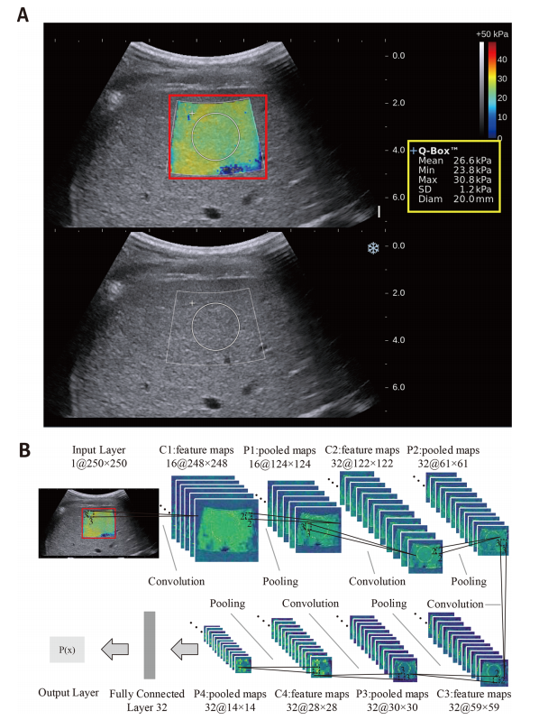
Figure 1 Illustration of the two-dimensional shear wave elastography (2D-SWE) measurement and the deep learning Radiomics of elastography (DLRE) flow chart. (A) The top shows the 2D-SWE region of interest (ROI) (pseudocolour area), Q-Box (white circle area within 2D-SWE ROI) and DLRE ROI (red square area). The obtained 2D-SWE values are displayed on the right yellow box. The bottom is the corresponding B-mode ultrasound image. (B) An input layer (DLRE ROI) is analysed by using four convolution-pooling procedures (C1-P1 to C4-P4), and then last pooled maps are fully connected with 32 neural nodes to calculate its probability for classification. The neural nodes and other parameters of the convolutional neural network (CNN) model were automatically optimised by using all 2D-SWE images in the training cohort.
图1 二维剪切波弹性成像(2D-SWE)测量和深度学习弹性成像放射组学(DLRE)流程图示例。(A) 顶部显示了2D-SWE感兴趣区域(ROI)(伪彩色区域)、Q-Box(2D-SWE ROI内的白色圆圈区域)和DLRE ROI(红色方形区域)。获得的2D-SWE值显示在右侧的黄色框中。底部是相应的B模式超声图像。(B) 输入层(DLRE ROI)通过四个卷积池化过程(C1-P1至C4-P4)进行分析,然后最后的池化图与32个神经节点完全连接,以计算其分类的概率。卷积神经网络(CNN)模型的神经节点和其他参数通过使用训练队列中的所有2D-SWE图像进行自动优化。
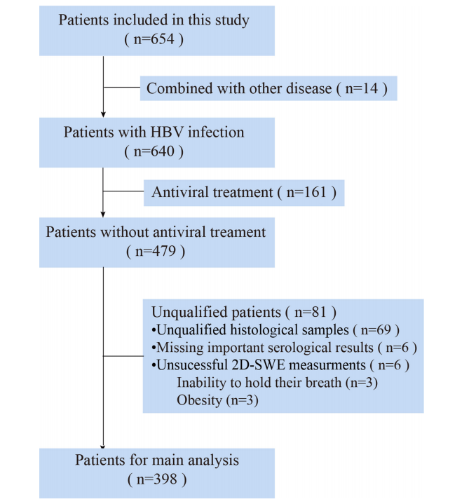
Figure 2 The results of the multicentre patient enrolments. In total, 398 out of 654 patients from 12 Chinese hospitals were enrolled in this study. 2D-SWE, two-dimensional shear wave elastography.
图2 多中心患者招募结果。总共,来自12家中国医院的654名患者中有398名被纳入了本研究。2D-SWE,二维剪切波弹性成像。
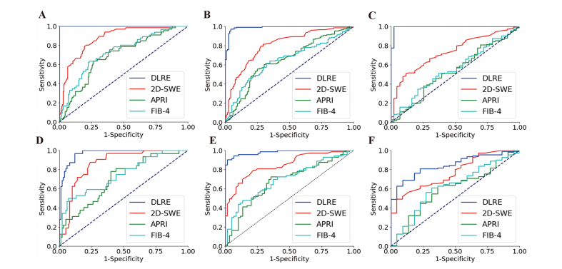
Figure 3 Comparison of ROC curves between DLRE, 2D-SWE and biomarkers for the assessment of liver fibrosis stages in training and validation cohorts, respectively. (A, D) F0-F3 versus F4 (F4) in training and validation cohorts. (B, E) F0-F2 versus F3-F4 (≥F3) in training and validation cohorts. (C, F) F0-F1 versus F2-F4 (≥F2) in training and validation cohorts. 2D-SWE, two-dimensional shear wave elastography; APRI, aspartate transaminaseto-platelet ratio index; DLRE, deep learning Radiomics of elastography; FIB-4, fibrosis index based on four factors.
图3 分别对比了DLRE、2D-SWE和生物标志物在训练队列和验证队列中评估肝纤维化阶段的ROC曲线。(A, D) 在训练和验证队列中,F0-F3与F4(F4)的对比。(B, E) 在训练和验证队列中,F0-F2与F3-F4(≥F3)的对比。(C, F) 在训练和验证队列中,F0-F1与F2-F4(≥F2)的对比。2D-SWE,二维剪切波弹性成像;APRI,天冬氨酸转氨酶至血小板比值指数;DLRE,弹性成像的深度学习放射组学;FIB-4,基于四个因素的纤维化指数。

Figure 4 Comparison of receiver operating characteristic (ROC) curves between deep learning Radiomics of elastography (DLRE) and twodimensional shear wave elastography (2D-SWE) using different number of image acquisitions/measurements (1, 3 and 5) of each patient for the assessment of liver fibrosis stages. (A, D) F0-F3 versus F4 (F4) in training and validation cohorts. (B, E) F0-F2 versus F3-F4 (≥F3) in training and validation cohorts. (C, F) F0-F1 versus F2-F4 (≥F2) in training and validation cohorts.
图4 深度学习弹性成像放射组学(DLRE)和二维剪切波弹性成像(2D-SWE)在每个患者的不同图像获取/测量数量(1、3和5)下对肝纤维化阶段进行评估的接收器操作特征(ROC)曲线比较。(A, D) 在训练和验证队列中,F0-F3与F4(F4)的对比。(B, E) 在训练和验证队列中,F0-F2与F3-F4(≥F3)的对比。(C, F) 在训练和验证队列中,F0-F1与F2-F4(≥F2)的对比。
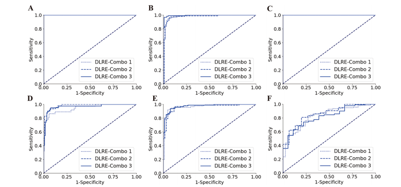
Figure 5 Comparison of receiver operating characteristic (ROC) curves between different combinations of hospitals for training deep learning Radiomics of elastography (DLRE) in the classification of liver fibrosis stages. (A, D) F0-F3 versus F4 (F4) in training (combination of hospitals B, D, G, E, H and J) and validation cohorts. (B, E) F0-F2 versus F3-F4 (≥F3) in training (combination of hospitals A, C and K) and validation cohorts. (C, F) F0-F1 versus F2-F4 (≥F2) in training (combination of hospitals A, G and K) and validation cohorts. Note: three ROC curves completely overlap each other in (A) and (C), as they all reach the optimal profile (area under the receiver operating characteristic curve (AUC)=1).
图5 在训练深度学习弹性成像放射组学(DLRE)用于肝纤维化阶段分类的不同医院组合之间的接收器操作特征(ROC)曲线比较。(A, D) 在训练队列(医院B、D、G、E、H和J的组合)和验证队列中,F0-F3与F4(F4)的对比。(B, E) 在训练队列(医院A、C和K的组合)和验证队列中,F0-F2与F3-F4(≥F3)的对比。(C, F) 在训练队列(医院A、G和K的组合)和验证队列中,F0-F1与F2-F4(≥F2)的对比。注意:在(A)和(C)中,三条ROC曲线完全重叠在一起,因为它们都达到了最佳性能(接收器操作特征曲线下面积(AUC)=1)。
Table
表
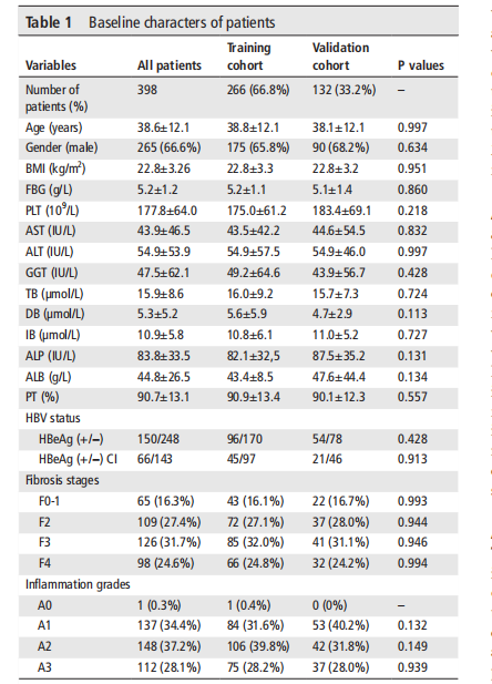
Table 1 Baseline characters of patients
表1 患者基线特征
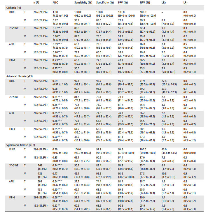
Table 2 Diagnostic performance of DLRE, 2D-SWE, APRI and FIB-4 for the assessment of liver fibrosis stages in training and validation cohorts
表2 DLRE、2D-SWE、APRI和FIB-4在训练队列和验证队列中评估肝纤维化阶段的诊断性能
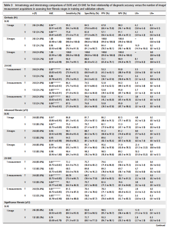
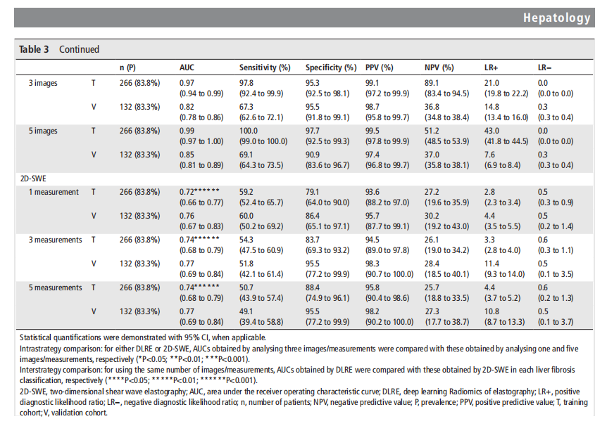
Table 3 Intrastrategy and interstrategy comparisons of DLRE and 2D-SWE for their relationship of diagnostic accuracy versus the number of image/measurement acquisitions in assessing liver fibrosis stages in training and validation cohorts
表3 DLRE和2D-SWE在训练队列和验证队列中对于图像/测量获取数量与评估肝纤维化阶段的诊断准确性关系的内部策略和策略间比较
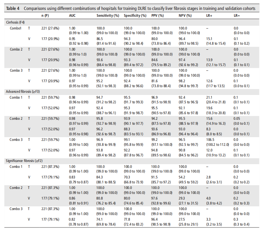
Table 4 Comparisons using different combinations of hospitals for training DLRE to classify liver fibrosis stages in training and validation cohorts
表4 使用不同医院组合对DLRE进行训练以对训练和验证队列中的肝纤维化阶段进行分类的比较


 678
678

 被折叠的 条评论
为什么被折叠?
被折叠的 条评论
为什么被折叠?
 到【灌水乐园】发言


